Review
. 2018 Jul;66(7):896-908.
doi: 10.4103/ijo.IJO_1136_17.

Polypoidal choroidal vasculopathy: Pearls in diagnosis and management

Affiliations
Review

Polypoidal choroidal vasculopathy: Pearls in diagnosis and management

Giridhar Anantharaman et al. Indian J Ophthalmol. 2018 Jul.

Abstract

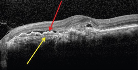
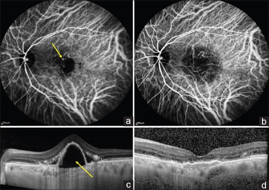
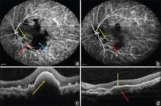
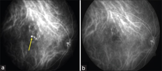
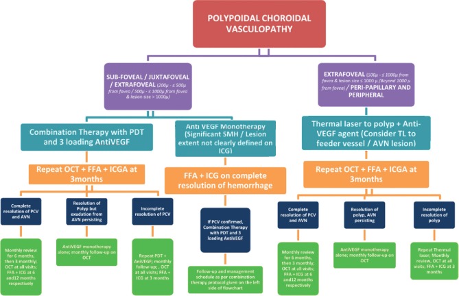
Polypoidal choroidal vasculopathy (PCV) is increasingly recognized as an important cause of exudative maculopathy in Asians as against Wet age-related macular degeneration in Caucasians. A panel of retinal experts methodically evaluated pertinent updated literature on PCV with thorough PubMed/MEDLINE search. Based on this, the panel agreed upon and proposed the current consensus recommendations in the diagnosis (clinical and imaging), management and follow-up schedule of PCV. Diagnosis of PCV should be based on the gold standard indocyanine green angiography which demonstrates early nodular hyperfluorescence signifying the polyp with additional features such as abnormal vascular network (AVN). Optical coherence tomography is an excellent adjuvant for diagnosing PCV, monitoring disease activity, and decision-making regarding the treatment. Current treatment modalities for PCV include photodynamic therapy, anti-vascular endothelial growth factor agents, and thermal laser. Choice of specific treatment modality and prognosis depends on multiple factors such as the location and size of PCV lesion, presence or absence of polyp with residual AVN, amount of submacular hemorrhage, presence or absence of leakage on fundus fluorescein angiography, visual acuity, and so on. Current recommendations would be invaluable for the treating physician in diagnosing PCV and in formulating the best possible individualized treatment strategy for optimal outcomes in PCV management.

Keywords: Abnormal vascular network; indocyanine green angiography; optical coherence tomography; photodynamic therapy; polypoidal choroidal vasculopathy; thermal laser.


Conflict of interest statement

There are no conflicts of interest

Figures

Figure 1
Figure 1
Color fundus photograph showing clinical types of polypoidal choroidal vasculopathy: (a) Exudative; (b) Hemorrhagic; (c) Mixed
Figure 2
Figure 2
Color fundus photograph (a) of a patient with polypoidal choroidal vasculopathy. Midphase indocyanine green angiography (b) showing the presence of polyp (Red arrow) and multifocal areas of hyperfluorescence (Yellow arrows)
Figure 3
Figure 3
Spectral domain optical coherence tomography images showing characteristic features of polypoidal choroidal vasculopathy: (a) Sharp-peaked pigment epithelial detachments (Yellow arrow); (b) Thumb-like pigment epithelial detachments, also called thumb-like polyp (thumb-like polyp; Yellow arrow); (c) Notch present at nasal portion of pigment epithelial detachments (Notched PED; Yellow arrow), along with hyporeflective lumen (Red arrow) surrounded by hyperreflective ring (Blue arrow) attached to undersurface of retinal pigment epithelium
Figure 4
Figure 4
Color fundus photograph (a) of a patient with serosanguinous polypoidal choroidal vasculopathy. Early phase indocyanine green angiography (b) showing the presence of abnormal vascular network (Yellow circle) in the absence of feeder vessel suggestive of interconnecting channels
Figure 5
Figure 5
Early-phase indocyanine green angiography showing the presence of a feeder vessel (Yellow arrow in a) with filling of the entire abnormal vascular network (Yellow arrows in b) suggestive of branch vascular network
Figure 6
Figure 6
Indocyanine green angiography images showing various topographic locations of polyps (Yellow arrow): (a) Subfoveal; (b) Juxtafoveal; (c) Extrafoveal; (d) Peripapillary; (e) Peripheral
Figure 7
Figure 7
Indocyanine green angiography image showing the presence of hyperfluorescent lesion with clearly demarcated geographic margin (Yellow arrows) suggestive of late geographic hyperfluorescence
Figure 8
Figure 8
Indocyanine green angiography showing the presence of multiple polyps with network of vessels in the foveal region. Yellow circle represents the total area (Polyp + abnormal vascular network) of polypoidal choroidal vasculopathy to be treated
Figure 9
Figure 9
Spectral domain optical coherence tomography showing the presence of double-layer sign, represented by two hyperreflective lines, inner signifying shallow retinal pigment epithelium elevation (Red arrow), and outer signifying Bruch's membrane (Yellow arrow) indicative of abnormal vascular network
Figure 10
Figure 10
Pretreatment image of a patient with nodular hyperfluorescence on indocyanine green angiography (a; Yellow arrow) and thumb-like pigment epithelial detachments on spectral domain optical coherence tomography (c; Yellow arrow). After 3 months of treatment with full-fluence photodynamic therapy and three doses of intravitreal ranibizumab, there was complete resolution of polypoidal choroidal vasculopathy on indocyanine green angiography (b; absence of nodular hyperfluorescence) and spectral domain optical coherence tomography (d; resolution of pigment epithelial detachments and fluid)
Figure 11
Figure 11
Pretreatment indocyanine green angiography (a) image of a patient with nodular hyperfluorescence (a; red arrow), abnormal vascular network (AVN; Yellow arrow), and large area of blocked hyperfluorescence due to hemorrhage (a; Blue arrow) and corresponding spectral domain optical coherence tomography (c) showing thumb-like pigment epithelial detachments with dense hyperreflectivity with backshadowing suggestive of hemorrhagic pigment epithelial detachments (Yellow arrow). After treatment with four doses of monthly intravitreal ranibizumab, indocyanine green angiography (b) showed residual polyps (b; Red arrow) and abnormal vascular network (b; Yellow arrow) while the blocked fluorescence resolved completely and corresponding spectral domain optical coherence tomography (d) demonstrated resolution of pigment epithelial detachments but persistence of double-layer sign represented by irregular retinal pigment epithelium elevation (d; Yellow line) and Bruch's membrane (d; Red arrow)
Figure 12
Figure 12
Pretreatment indocyanine green angiography (a) demonstrating nodular hyperfluorescence (Yellow arrow) suggestive of extrafoveal polypoidal choroidal vasculopathy. After treatment with thermal laser and three loading doses of intravitreal ranibizumab, there was complete resolution of polyps on indocyanine green angiography (b)
Figure 13
Figure 13
Flowchart for management of PCV based on the updated guidelines

Similar articles

Cited by

References

    1. Yannuzzi LA. Miami, FL: 1982. Idiopathic polypoidal choroidal vasculopathy. Macula Society Meeting.
    1. Liu Y, Wen F, Huang S, Luo G, Yan H, Sun Z, et al. Subtype lesions of neovascular age-related macular degeneration in chinese patients. Graefes Arch Clin Exp Ophthalmol. 2007;245:1441–5. - PubMed
    1. Maruko I, Iida T, Saito M, Nagayama D, Saito K. Clinical characteristics of exudative age-related macular degeneration in Japanese patients. Am J Ophthalmol. 2007;144:15–22. - PubMed
    1. Chang YC, Wu WC. Polypoidal choroidal vasculopathy in Taiwanese patients. Ophthalmic Surg Lasers Imaging. 2009;40:576–81. - PubMed
    1. Byeon SH, Lee SC, Oh HS, Kim SS, Koh HJ, Kwon OW, et al. Incidence and clinical patterns of polypoidal choroidal vasculopathy in Korean patients. Jpn J Ophthalmol. 2008;52:57–62. - PubMed