Review
. 2018 Jul;66(7):909-915.
doi: 10.4103/ijo.IJO_136_18.

Novel techniques in scleral buckling

Affiliations
Review

Novel techniques in scleral buckling

P Mahesh Shanmugam et al. Indian J Ophthalmol. 2018 Jul.

Abstract

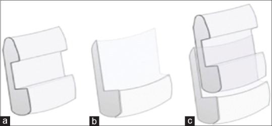
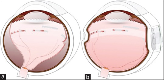
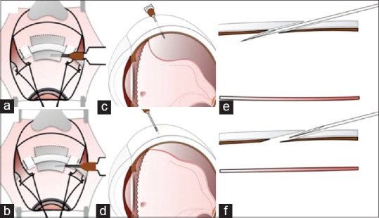
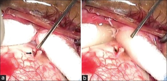
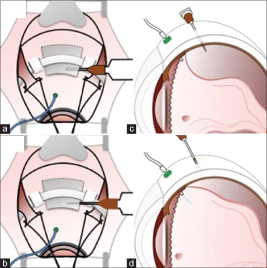
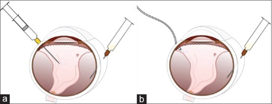
Scleral buckling is a surgical technique that is employed successfully to treat rhegmatogenous retinal detachments (RRD) for more than 60 years. With the introduction of pars plana vitrectomy (PPV), there is a growing trend towards the use of PPV for treatment of retinal detachment. There is a reluctance to perform scleral buckling (SB) in RRD due to the perceived steep learning curve, declining mastery over indirect ophthalmoscopy, and poor ergonomics associated with SB. In this article, we discuss the surgical challenges and tips to overcome these in four headings: localization of the break, retinopexy, SB, and subretinal fluid (SRF) drainage. Localization of the break can be performed by the use of forceps or illuminated scleral depressor. It can be facilitated by prior drainage of SRF in cases with bullous RRD. Chandelier with wide-angle viewing system can be used for easier localization of break and cryopexy. Sutureless buckling and suprachoroidal buckling are easier and faster alternatives to the conventional technique. Reshaping the silicone segment helps in accommodating the wider circumferential band. Modified needle drainage, laser choroidotomy, and infusion-assisted drainage can make SRF drainage easier and safer. The above techniques and other practical tips have been explained in detail with the illustrations to make the process of learning the art of SB easier.

Keywords: Retinal detachment; retinopexy; scleral buckling; subretinal fluid drainage.


Conflict of interest statement

There are no conflicts of interest

Figures

Figure 1
Figure 1
Localization of break with forceps
Figure 2
Figure 2
Localization of break in chandelier-assisted surgery. (a) Break is seen (white arrow) at the summit of indent through the wide-angle viewing system. (b) Grey dot on sclera (black arrow) due to indentation by forceps tip corresponding to the break
Figure 3
Figure 3
Illuminated scleral indenter. (a) The tip of the light pipe (preferably 20G) is bent. (b) View through 20D lens. The glow of light pipe tip (white arrow) seen on the retina adjacent to the lateral edge of tear (black arrow) by trans-scleral illumination
Figure 4
Figure 4
Location of the break with cryoprobe
Figure 5
Figure 5
Cryotherapy in chandelier-assisted surgery. Cryo edema is seen around the horse shoe tear through the wide angle viewing system
Figure 6
Figure 6
(a) Illuminated cryoprobe. (b) View through 20D lens. Cryo edema around the horse shoe tear. Red glow is due to the illuminated tip
Figure 7
Figure 7
(a) Retinal detachment with multiple lattices with holes and absence of vitreous detachment (green). (b) 4 mm band to support the posterior most lesion. Vitreous plugs the holes and allows retinal reattachment
Figure 8
Figure 8
Suture less scleral buckling. Buckle is placed in between the two scleral tunnels (black arrow) and supported by encerclage
Figure 9
Figure 9
(a) Increasing the width of the groove on the buckle. (b) Suture less buckle placed beneath 4 mm band
Figure 10
Figure 10
Intrascleral buckle. (a) Circumferential partial thickness scleral incision over the break. (b) Scleral pocket is created anteriorly and posteriorly. (c) The silicone segment is placed in the scleral pocket. (d) Suture is used to close the pocket
Figure 11
Figure 11
Suprachoroidal buckling. (a) A radial incision is placed at a distance from the localization mark. (b) The choroid is separated gently at the site of sclerotomy by injecting viscoelastic. (c) Cannula is threaded in the suprachoroidal space to reach the mark. (d) Viscoelastic is injected in the suprachoroidal space at the site of break. Laser photocoagulation is performed around the break with laser indirect ophthalmoscope
Figure 12
Figure 12
Suprachoroidal buckling. (a) Radial incision made at a distance from the mark so that the tip of metal cannula can reach the mark. (b) Choroid is separated gently at the site of sclerotomy. (c) Cannula is threaded in the suprachoroidal space to reach the mark
Figure 13
Figure 13
(a) Silicone segment. (b) Superficial part of the segment anterior to the groove is shaved off. (c) The modified buckle is placed beneath another buckle
Figure 14
Figure 14
(a) Posterior tear with extensive lattices with holes. (b) Tear supported by the technique described in Figure 13
Figure 15
Figure 15
Modifications of needle drainage. (a, c and e) Needle is inserted obliquely with the bevel facing upward (toward the choroid, e). (b, d, and f) Needle is retracted back partially such that the bevel is retained within the track
Figure 16
Figure 16
Laser choroidotomy. (a) After the scleral cut down. The aiming beam (red dot indicated by the arrow) of laser from the endolaser probe is directed to the choroid. (b) Successful drainage of subretinal fluid after the laser shot
Figure 17
Figure 17
Infusion-assisted drainage in bullous retinal detachment. Infusion cannula is placed in the same quadrant of drainage. (a and c) Needle is inserted obliquely with the bevel facing upwards. (b and d) Needle is retracted back partially. Subretinal fluid starts draining and simultaneous infusion combats hypotony
Figure 18
Figure 18
Needle drainage under visualization. 26G needle loaded on to a syringe (with plunger removed) is obliquely inserted in the eye under visualization with indirect ophthalmoscope. (a) Assistant aided injection of balanced salt solution to combat hypotony. (b) Simultaneous infusion to combat hypotony

Similar articles

Cited by

References

    1. Park SW, Kwon HJ, Byon IS, Lee JE, Oum BS. Impact of age on scleral buckling surgery for rhegmatogenous retinal detachment. Korean J Ophthalmol. 2017;31:328–35. - PMC - PubMed
    1. Feng H, Adelman RA. Cataract formation following vitreoretinal procedures. Clin Ophthalmol. 2014;8:1957–65. - PMC - PubMed
    1. Blodi BA, Paluska SA. Cataract after vitrectomy in young patients. Ophthalmology. 1997;104:1092–5. - PubMed
    1. Hotta K, Sugitani A. Refractive changes in silicone oil-filled pseudophakic eyes. Retina. 2005;25:167–70. - PubMed
    1. Stefánsson E, Anderson MM, Jr, Landers MB, 3rd, Tiedeman JS, McCuen BW., 2nd Refractive changes from use of silicone oil in vitreous surgery. Retina. 1988;8:20–3. - PubMed