. 2018 Jun 25;8(7):117.
doi: 10.3390/brainsci8070117.

Pallidal Stimulation Modulates Pedunculopontine Nuclei in Parkinson's Disease

Affiliations

Pallidal Stimulation Modulates Pedunculopontine Nuclei in Parkinson's Disease

Imke Galazky et al. Brain Sci. .

Abstract

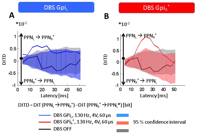
Background: In advanced Parkinson’s disease, the pedunculopontine nucleus region is thought to be abnormally inhibited by gamma-aminobutyric acid (GABA) ergic inputs from the over-active globus pallidus internus. Recent attempts to boost pedunculopontine nucleus function through deep brain stimulation are promising, but suffer from the incomplete understanding of the physiology of the pedunculopontine nucleus region.

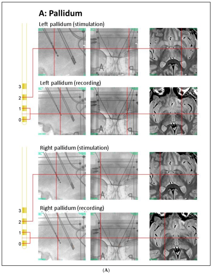
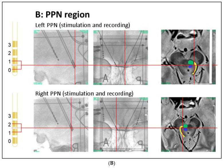
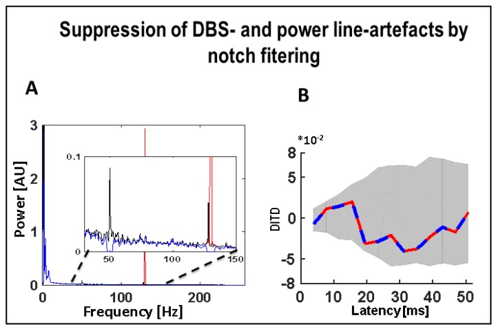
Methods: Local field potentials of the pedunculopontine nucleus region and the globus pallidus internus were recorded and quantitatively analyzed in a patient with Parkinson’s disease. In particular, we compared the local field potentials from the pedunculopontine nucleus region at rest and during deep brain stimulation of the globus pallidus internus.

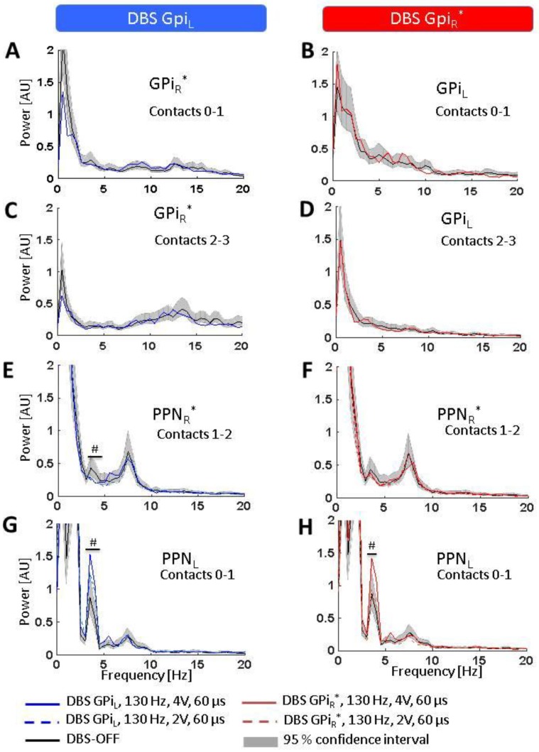
Results: At rest, the spectrum of local field potentials in the globus pallidus internus was mainly characterized by delta-theta and beta frequency activity whereas the spectrum of the pedunculopontine nucleus region was dominated by activity only in the delta and theta band. High-frequency deep brain stimulation of the globus pallidus internus led to increased theta activity in the pedunculopontine nucleus region and enabled information exchange between the left and right pedunculopontine nuclei. Therefore, Conclusions: When applying deep brain stimulation in the globus pallidus internus, its modulatory effect on pedunculopontine nucleus physiology should be taken into account.

Keywords: Parkinson’s disease; deep brain stimulation; globus pallidus internus; local field potentials; pedunculopontine nucleus.


Conflict of interest statement

The authors declare no conflicts of interest.

Figures

Figure 1
Figure 1
Anatomical positions of DBS electrodes. (A) Anatomical location of the GPi electrodes as determined by intra-operative orthogonal x-ray imaging: in the lateral and frontal x-ray views, the location of each contact used for stimulation or recording is marked by a red cross, as is the corresponding location in the proton weighted MRI (from left to right). Orthogonal x-ray images and intra-operative CT were acquired with stereotactic image fiducials allowing for transformation into a common (stereotactic) coordinate system with a spatial mismatch between CT and x-ray coordinates of less than 1 mm (unpublished data). MRI images were transformed into this coordinate system by manual image registration to the CT data using anatomical landmarks. Contacts 0–2 of the left electrode and contacts 0 and 1 of the right electrode were located within the ventro–postero–lateral section of the respective GPi. Please note that LFP signals were recorded and analyzed both from electrode pairs 0–1 and 2–3 (during contralateral stimulation); (B) Anatomical location of the PPN electrodes: the PPN region is located lateral to the decussation of the superior cerebellar peduncles (green) and the central segmental tract (blue) and medial to the lemniscal systems (yellow) (see Zrinzo et al., 2008 for anatomical details). According to these landmarks, contacts 0–2 of the left electrode and contacts 1 and 2 of the right electrode were located within the respective rostral PPN. Abbreviations: CT: computer tomography, DBS: deep brain stimulation, GPi: globus pallidus internus, MRI: magnetic resonance imaging, PPN: pedunculopontine nucleus region.
Figure 1
Figure 1
Anatomical positions of DBS electrodes. (A) Anatomical location of the GPi electrodes as determined by intra-operative orthogonal x-ray imaging: in the lateral and frontal x-ray views, the location of each contact used for stimulation or recording is marked by a red cross, as is the corresponding location in the proton weighted MRI (from left to right). Orthogonal x-ray images and intra-operative CT were acquired with stereotactic image fiducials allowing for transformation into a common (stereotactic) coordinate system with a spatial mismatch between CT and x-ray coordinates of less than 1 mm (unpublished data). MRI images were transformed into this coordinate system by manual image registration to the CT data using anatomical landmarks. Contacts 0–2 of the left electrode and contacts 0 and 1 of the right electrode were located within the ventro–postero–lateral section of the respective GPi. Please note that LFP signals were recorded and analyzed both from electrode pairs 0–1 and 2–3 (during contralateral stimulation); (B) Anatomical location of the PPN electrodes: the PPN region is located lateral to the decussation of the superior cerebellar peduncles (green) and the central segmental tract (blue) and medial to the lemniscal systems (yellow) (see Zrinzo et al., 2008 for anatomical details). According to these landmarks, contacts 0–2 of the left electrode and contacts 1 and 2 of the right electrode were located within the respective rostral PPN. Abbreviations: CT: computer tomography, DBS: deep brain stimulation, GPi: globus pallidus internus, MRI: magnetic resonance imaging, PPN: pedunculopontine nucleus region.
Figure 2
Figure 2
Effect of notch filters to suppress power line and DBS related artefacts. (A) Power spectrum of left PPN-LFP at base line (black line) with a simulated strong 130 Hz artefact (red line) superimposed. After notch filtering at 50 Hz and 130 Hz, the corresponding components are suppressed (blue line), the additional peak at 128 reflects environmental noise generated by surrounding medical equipment; (B) The difference of the DIT in the two directions (i.e., DITD = DIT (PPNL → PPNR*) − DIT (PPNR* → PPNL*)). Blue: DITD between the PPN-LFPs (without the simulated 130 Hz artefact) after notch filtering (50 Hz, 130 Hz). Red: DITD between the PPN-LFPs (both including superimposed simulated 130 Hz artefacts of equal strength) after notch filtering (50 Hz, 130 Hz). Abbreviations: DBS: deep brain stimulation, DIT: directed information transfer, LFP: local field potentials, PPN: pedunculopontine nucleus region (L = left, R = right, * denotes clinically more affected side).
Figure 3
Figure 3
Effects of GPi-DBS on LFP spectra. Statistical significance levels of differences were derived by comparing the DBS on-spectra to the chi-square distribution assumed for the base line (DBS-OFF) spectra (see Welch, 1967). Significant differences are marked by a ‘#’ indicating a significance level of p < 0.05. (AD) No spectral changes were observed in GPiR* or GPiL (neither at contacts 0–1 nor 2–3) during DBS of the contralateral GPi; (E,F) A slight but significant decrease was observed in the lower theta band in PPNR* during GPIL-DBS but no spectral change was observed in PPNR* during GPiR*-DBS; (G,H) DBS of either GPiL (left) or GPiR* (right) led to increased activity in the lower theta band in PPNL. Abbreviations: DBS: deep brain stimulation, GPi: globus pallidus internus (L = left, R = right, * denotes clinically more affected side), LFP: local field potentials, PPN: pedunculopontine nucleus region (L = left, R = right, * denotes clinically more affected side).
Figure 4
Figure 4
Effects of GPi-DBS on information exchange. Data are presented as differences in directed information transfer (DIT), i.e., values greater or smaller than zero indicate opposite directions of information flow as indicated on the diagrams, and DIT magnitude gives the amount of information exchanged in bits. Shaded areas give the 95% confidence interval of the DIT estimates after time-shuffling one of the two signals (leading to zero coupling), i.e., only DIT values outside the CI can be considered significantly different from zero. (A,B) While no exchange of information is observed between both PPNs during DBS-OFF, DBS of either GPiL or GPiR* induces a significant flow of information directed from PPNL to PPNR*. Abbreviations: CI: confidence interval, DBS: deep brain stimulation, DIT: directed information transfer, GPi: globus pallidus internus (L = left, R = right, *denotes clinically more affected side), PPN: pedunculopontine nucleus region (L = left, R = right, *denotes clinically more affected side).

Similar articles

Cited by

References

    1. Rahman S., Griffin H.J., Quinn N.P., Jahanshahi M. Quality of life in Parkinson’s disease: The relative importance of the symptoms. Mov. Disord. 2008;23:1428–1434. doi: 10.1002/mds.21667. - DOI - PubMed
    1. St George R.J., Nutt J.G., Burchiel K.J., Horak F.B. A meta-regression of the long-term effects of deep brain stimulation on balance and gait in PD. Neurology. 2010;75:1292–1299. doi: 10.1212/WNL.0b013e3181f61329. - DOI - PMC - PubMed
    1. Benarroch E.E. Pedunculopontine nucleus: Functional organization and clinical implications. Neurology. 2013;80:1148–1155. doi: 10.1212/WNL.0b013e3182886a76. - DOI - PubMed
    1. Stefani A., Lozano A.M., Peppe A., Stanzione P., Galati S., Tropepi D., Pierantozzi M., Brusa L., Scarnati E., Mazzone P. Bilateral deep brain stimulation of the pedunculopontine and subthalamic nuclei in severe Parkinson’s disease. Brain J. Neurol. 2007;130:1596–1607. doi: 10.1093/brain/awl346. - DOI - PubMed
    1. Mazzone P., Insola A., Sposato S., Scarnati E. The deep brain stimulation of the pedunculopontine tegmental nucleus. Neuromodul. J. Int. Neuromodul. Soc. 2009;12:191–204. doi: 10.1111/j.1525-1403.2009.00214.x. - DOI - PubMed

LinkOut - more resources