. 2018 Jun 25;8(1):9633.
doi: 10.1038/s41598-018-27890-4.

Characterization of influenza virus variants induced by treatment with the endonuclease inhibitor baloxavir marboxil

Affiliations

Characterization of influenza virus variants induced by treatment with the endonuclease inhibitor baloxavir marboxil

Shinya Omoto et al. Sci Rep. .

Abstract

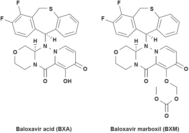
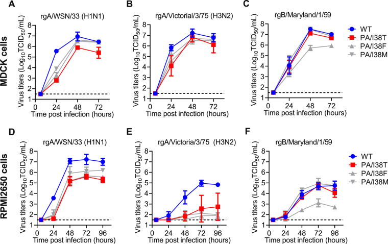
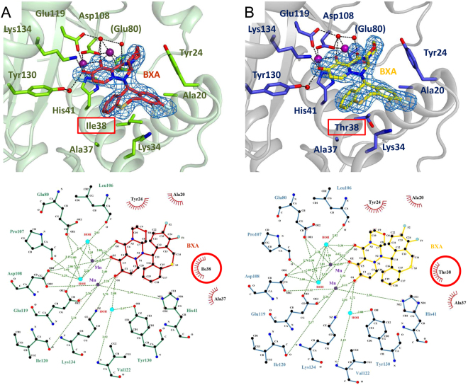
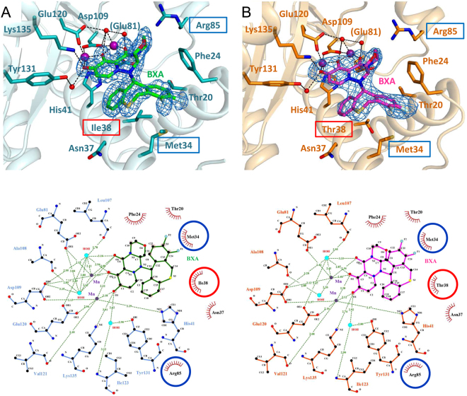
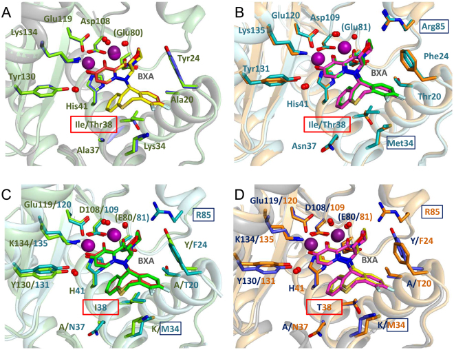
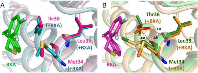
Baloxavir acid (BXA), derived from the prodrug baloxavir marboxil (BXM), potently and selectively inhibits the cap-dependent endonuclease within the polymerase PA subunit of influenza A and B viruses. In clinical trials, single doses of BXM profoundly decrease viral titers as well as alleviating influenza symptoms. Here, we characterize the impact on BXA susceptibility and replicative capacity of variant viruses detected in the post-treatment monitoring of the clinical studies. We find that the PA I38T substitution is a major pathway for reduced susceptibility to BXA, with 30- to 50-fold and 7-fold EC50 changes in A and B viruses, respectively. The viruses harboring the I38T substitution show severely impaired replicative fitness in cells, and correspondingly reduced endonuclease activity in vitro. Co-crystal structures of wild-type and I38T influenza A and B endonucleases bound to BXA show that the mutation reduces van der Waals contacts with the inhibitor. A reduced affinity to the I38T mutant is supported by the lower stability of the BXA-bound endonuclease. These mechanistic insights provide markers for future surveillance of treated populations.


Conflict of interest statement

All authors, except V.S. and S.C., were employees of Shionogi & Co, Ltd. in Osaka, Japan. The results in the paper were partially generated by the funding from Shionogi & Co., Ltd.

Figures

Figure 1
Figure 1
Chemical structure of baloxavir acid (BXA, active form) and baloxavir marboxil (prodrug form). The full chemical name of BXA is (12aR)-12-[(11 S)-7,8-difluoro-6,11-dihydrodibenzo[b,e] thiepin-11-yl]-7-hydroxy-3,4,12,12a-tetrahydro-1H-[1,4] oxazino[3,4-c]pyrido[2,1-f][1,2,4]triazine-6,8-dione.
Figure 2
Figure 2
Replicative capacity of variant viruses with indicated AA substitutions in PA protein. Canine MDCK cells (AC) or human RPMI2650 cells (D,E) were infected with WT or I38x viruses based on rgA/WSN/33 (H1N1) (A,D), rgA/Victoria/3/75 (H3N2) (B,E), or B/Maryland/1/59 (C,F). The culture supernatants were collected at the indicated time points and viral titers (TCID50/mL) were determined in MDCK cells. Each symbol represents the mean and standard deviation of triplicate experiments. The lower limit of quantification of the virus titers was indicated by a dashed line.
Figure 3
Figure 3
In vitro endonuclease activity and inhibition of PA variants and thermal stabilization induced by the binding of BXA. (A) For endonuclease activity (lane marked - in all panels), FAM-RNA was incubated with either 0.5–5 μM PA-A-WT, 5–50 μM PA-A-I38T, 5–50 µM PA-B-WT or 50–500 µM PA-B-I38T. Reactions were carried out for 2 hours in the presence of 2.5 mM MnCl2 at 37 °C and were stopped by adding 1 mM EDTA. For inhibition assays, all four proteins were pre-incubated with increasing concentrations of BXA for 30′ at RT and then tested for activity. In all cases, four compound concentrations were tested, maintaining the molar ratio to protein equal to 0.1–0.2–0.5–1 (in all panels, WT: lanes 4–7, I38T: lanes 9-12). The reaction products were loaded on an 8 M 20% acrylamide gel and detected at 520 nm using standard emission filter in Gel Doc™ XR+ (BioRad). Images were processed using Image Lab software (v 6.0.0.25, BioRad) as detailed in Methods. Gels were cropped (removal of gel wells and band-less edges) for clarity. Black triangles (▼) compare the RNase activity of WT and I38T forms at equal protein concentration for FluA (5 µM, left) and FluB (50 µM right). Red asterisks (*) indicate the BXA concentration at which RNase activity is significantly reduced or abolished. For all panels, lane 1 “M”: molecular ladder (FAM-RNAs; numbers represent the RNAs size in nt). Lane 2 “U”: uncleaved RNA, input substrate. (B) BXA binding stabilizes PA WT (blue, green) and PA I38T (red, purple) for both influenza A (left) and influenza B (right). Thermal shift assays were performed with 13 µM PA (purified with buffer supplemented with 2 mM MnCl2 and 2 mM MgCl2) in 20 mM Tris/HCl pH 7.5, 100 mM NaCl in the presence or absence of 0-1.3 mM of BXA and SYPRO Orange dye (Invitrogen). The curves represent the fluorescence emission of the dye at 575 nm during protein denaturation. Green and purple curves represent the stabilization effect induced by BXA at concentration ≥36 μM in complex with WT and I38T constructs, respectively. The estimated Tm values, taken as mid-points between local minima and maxima, are indicated and listed in Table 2.
Figure 4
Figure 4
BXA binding to influenza A/H1N1 PA endonuclease. BXA interacts with (A) PA-A WT and (B) PA-A I38T by chelating the two manganese ions in the active site. Top panels represent the 3D structure of BXA-bound PA-A endonuclease, whereas the bottom ones are the corresponding 2D interaction ligplots. In the structures, both BXA (in TV red for PA WT and yellow for PA I38T) and the interacting side chains (in chartreuse green for WT and TV blue for I38T) are represented as sticks, Mn ions (purple) and water molecules (red) as spheres. In blue mesh, the omit Fo-Fc density map for BXA at 3.0 σ. In the ligplots, dash lines represent hydrogen bonds, red crowns hydrophobic interactions and red circles highlight the mutation site at position 38. Structural figures were prepared using Pymol (www.pymol.org), ligand interaction plots with LigPlot+ (EMBL−EBI).
Figure 5
Figure 5
BXA binding to influenza B/Memphis PA endonuclease As Fig. 4, but for (A) PA-B WT and (B) PA-B I38T. Top panels: 3D structures of BXA-bound PA-B. Bottom panels: corresponding 2D interaction ligplots. Both BXA (in green for PA WT and light magenta for PA I38T) and the interacting side chains (in teal for WT and orange for I38T) are represented as sticks, Mn ions (purple) and water molecules (red) as spheres. The protein main chain is drawn as a cartoon. Represented by the blue mesh is the omit Fo-Fc density map for BXA at 3.0 σ. In the ligplots, dash lines represent hydrogen bonds and red crowns hydrophobic interactions. Red circles highlight the mutation site, blue circles the additional hydrophobic contacts that characterize binding of BXA to PA-B. Ligand interaction plots were prepared with LigPlot+ (EMBL-EBI).
Figure 6
Figure 6
Comparison of PA endonuclease from Flu A and Flu B bound to BXA in either WT or I38T form. Superposition of PA-BXA complexes: (A) PA-A WT and PA-A I38T, (B) PA-B WT and PA-B I38T, (C) PA-A WT and PA-B WT, (D) PA-A I38T and PA-B I38T. Complexes PA/BXA are represented in sticks and coloured in chartreuse/TV red for A WT, TV blue/yellow for A I38T, teal/green for B WT, orange/light magenta for B I38T. Manganese ions and water residues involved in protein-inhibitor interactions are depicted as spheres, in purple and red, respectively. Protein main chain is represented as a cartoon. Highlighted in red rectangles are the mutation sites. The binding of the inhibitor does not differ significantly among the four protein constructs, which share high conservation in the local network of interactions with BXA, except locally when residue 38 is mutated. Additional contacts in the case of Influenza B are shown in blue rectangles in B, C and D.
Figure 7
Figure 7
Local interactions of residue 38 in apo- and BXA-bound FluB PA (A) Superposition of ligand-free PA-B WT (PDB:5FML, in hotpink) and bound to BXA (green sticks for BXA, teal sticks/cartoon for PA). (B) Superposition of ligand-free (forest green) and BXA-bound PA-B I38T (light magenta sticks for BXA, orange sticks/cartoon for PA). For the WT, BXA binding does not alter the disposition of Ile38 and neighbouring Met34. For the I38T mutant, both side-chains change rotamer and the contacts with both the main chain and the compound hydrophobic pocket are altered.

References

    1. Thompson WW, et al. Estimates of US influenza-associated deaths made using four different methods. Influenza and other respiratory viruses. 2009;3:37–49. doi: 10.1111/j.1750-2659.2009.00073.x. - DOI - PMC - PubMed
    1. Lambert LC, Fauci AS. Influenza vaccines for the future. The New England journal of medicine. 2010;363:2036–2044. doi: 10.1056/NEJMra1002842. - DOI - PubMed
    1. Dong G, et al. Adamantane-resistant influenza a viruses in the world (1902–2013): frequency and distribution of M2 gene mutations. PloS one. 2015;10:e0119115. doi: 10.1371/journal.pone.0119115. - DOI - PMC - PubMed
    1. Treanor JJ, et al. Efficacy and safety of the oral neuraminidase inhibitor oseltamivir in treating acute influenza: a randomized controlled trial. US Oral Neuraminidase Study Group. Jama. 2000;283:1016–1024. doi: 10.1001/jama.283.8.1016. - DOI - PubMed
    1. Nicholson KG, et al. Efficacy and safety of oseltamivir in treatment of acute influenza: a randomised controlled trial. Neuraminidase Inhibitor Flu Treatment Investigator Group. Lancet. 2000;355:1845–1850. doi: 10.1016/S0140-6736(00)02288-1. - DOI - PubMed

Publication types

MeSH terms