. 2018 Jun 25;9(1):2456.
doi: 10.1038/s41467-018-04957-4.

Site-specific characterization of endogenous SUMOylation across species and organs

Affiliations

Site-specific characterization of endogenous SUMOylation across species and organs

Ivo A Hendriks et al. Nat Commun. .

Abstract

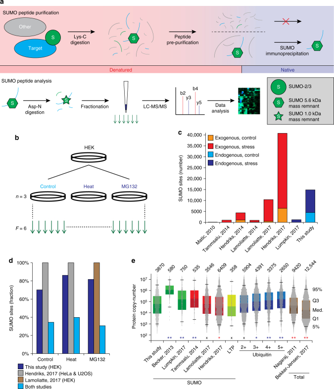
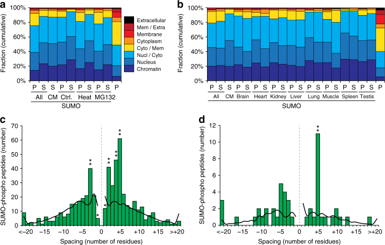
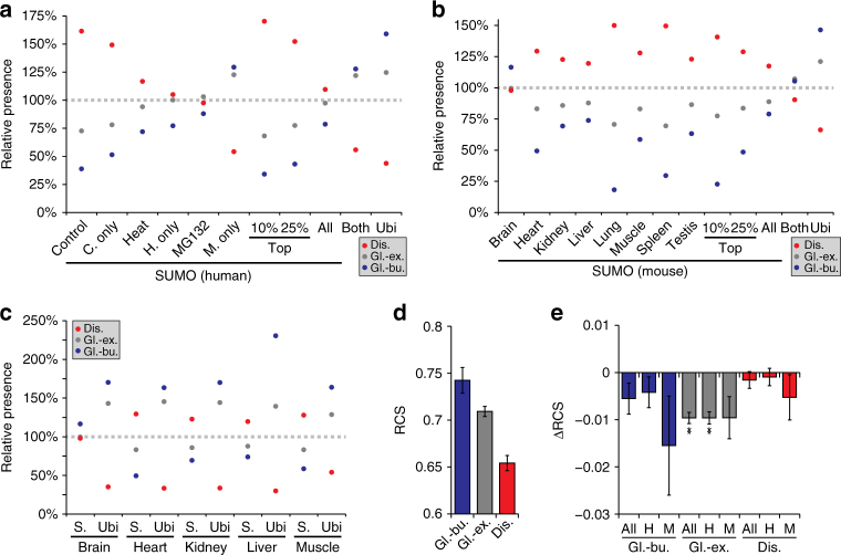
Small ubiquitin-like modifiers (SUMOs) are post-translational modifications that play crucial roles in most cellular processes. While methods exist to study exogenous SUMOylation, large-scale characterization of endogenous SUMO2/3 has remained technically daunting. Here, we describe a proteomics approach facilitating system-wide and in vivo identification of lysines modified by endogenous and native SUMO2. Using a peptide-level immunoprecipitation enrichment strategy, we identify 14,869 endogenous SUMO2/3 sites in human cells during heat stress and proteasomal inhibition, and quantitatively map 1963 SUMO sites across eight mouse tissues. Characterization of the SUMO equilibrium highlights striking differences in SUMO metabolism between cultured cancer cells and normal tissues. Targeting preferences of SUMO2/3 vary across different organ types, coinciding with markedly differential SUMOylation states of all enzymes involved in the SUMO conjugation cascade. Collectively, our systemic investigation details the SUMOylation architecture across species and organs and provides a resource of endogenous SUMOylation sites on factors important in organ-specific functions.


Conflict of interest statement

The authors declare no competing interests.

Figures

Fig. 1
Fig. 1
A strategy for identifying endogenous SUMO2/3 sites. a Schematic overview of the purification strategy. Briefly, a denaturing lysate is prepared and digested with Lys-C. Peptides are pre-purified using C8 SepPak cartridges, after which peptides are lyophilized. The peptides are then dissolved in a mild buffer to facilitate immunoprecipitation using the 8A2 antibody. Purified SUMOylated peptides are subjected to a second round of digestion using Asp-N, after which they are fractionated on StageTip and analyzed by nanoscale LC-MS/MS. b Experimental design for the main cell culture data. All experiments were performed in cell culture triplicates, and analyzed as six fractions. c Overview of the total number of SUMO2/3 sites identified in this study, as compared to several other SUMO proteomics studies. d Overview of the relative number of SUMO2/3 sites identified per condition in this study, as compared to the largest currently published SUMO proteomics studies that used comparable cellular treatments,. e Estimation of depth of sequencing through comparison of the copy-number per cell of identified proteins. IBAQ-determined copy-numbers of proteins were derived from a recent deep total HeLa proteome study. Protein copy-numbers were assumed to be comparable on average across different cultured human cell lines. Top whisker: 95th percentile, top bound: 3rd quantile, center line: median, bottom bound: 1st quantile, bottom whisker: 5th percentile. Total numbers of proteins identified are displayed above the bars. The copy-number distribution histogram of detected proteins is overlaid for all datasets. For ubiquitin, the numbers indicate the tested number of ubiquitylated lysines per protein, as extracted from the PhosphoSitePlus ubiquitin site database. LTP low throughput. Asterisks denote significant differences between SUMO2/3 target proteins identified in this study and other datasets, with blue or red asterisks indicating our study achieved more or less depth, respectively. Determined by two-tailed Fisher’s exact testing. * P < 0.05, ** P < 0.001, x N.S
Fig. 2
Fig. 2
The endogenous SUMO2/3 consensus motif and SUMO dynamics. a IceLogo representation of amino acid residues enriched around the top 10% SUMOylated lysines identified in human cells, at position 0, indicated by the asterisk. Residues displayed above the line are enriched, and those displayed below the line are depleted, compared to a reference set of human nuclear proteins. b The adherence of all identified SUMO2/3 sites to the KxE motif, in relation to the average of top-scoring sites. c Venn diagram of SUMO2/3 sites identified in response to different treatments, drawn to scale. d Venn diagram of SUMO2/3 target proteins identified in response to different treatments, drawn to scale
Fig. 3
Fig. 3
Mapping the mouse SUMOylome. a Experimental design of SUMO proteomics experiments carried out in mice. Eight types of organs were studied, in biological quintuplicate (five animals), and analyzed as six fractions. b Overview of the number of SUMO2/3 sites identified per organ, comparing direct MS/MS identifications, to identifications made when matching MS1-level data between organs. c Overview of the number of organs in which the same SUMO2/3 sites were detected. d The adherence of all identified SUMO2/3 sites to the KxE motif, in relation to the average of top-scoring sites. e IceLogo representation of amino acid residues enriched around all SUMOylated lysines identified in mouse, at position 0, indicated by the asterisk. Residues displayed above the line are enriched, and those displayed below the line are depleted, compared to a reference set of mouse nuclear proteins. f The adherence of MS/MS-identified SUMO2/3 sites to the KxE motif, per organ, relative to the normalized expected adherence based on (d). g Term enrichment analysis comparing identified mouse SUMO2/3 target proteins to mouse proteins known to be expressed in the eight organs analyzed. Numbers in parentheses indicate overlap between the numbers of proteins identified in this study, compared to those present in the reference. Score was derived from the logarithms of enrichment ratios and Benjamini–Hochberg FDR-corrected two-tailed Fisher’s P-values of < 0.02. A full list of all enriched terms, and all relevant scores, is available in Supplementary Data 7
Fig. 4
Fig. 4
Analysis of SUMOylation across mouse organs. a Hierarchical clustering analysis of Z-scored label-free quantified (LFQ) expression values corresponding to mouse SUMO2/3 target proteins detected across replicates and organs. Blue coloring indicates relative presence in a sample as compared to others. b Principle component analysis (PCA) of all mouse experiments. The principle components represent the greatest degree of variability observed within the data, and grouped experiments are generally more similar than distant experiments. Eigenvalues are displayed on the axes. c Qualitative term enrichment analysis comparing SUMO2/3 target proteins between organs, with all SUMO target proteins inferred from MS/MS-identified SUMO site. SUMO2/3 target proteins were compared against corresponding organ background proteomes, and only terms that were significantly enriched are displayed. Black text indicates terms which additionally correspond to functions that were uniquely found to be enriched in one organ, whereas blue text indicates terms that were enriched in more than one organ. Numbers in parentheses indicate overlap between the numbers of SUMO target proteins identified in the organ, compared to those present in the reference. Score was derived from the logarithms of enrichment ratios and Benjamini-Hochberg FDR-corrected two-tailed Fisher’s P-values of <0.02. A full list of all enriched terms, and all relevant scores, is available in Supplementary Data 8. d Overview of the relative expression levels of SUMO2/3 target proteins identified in each mouse organ, as compared to the expression levels of the same proteins in the other organs. The dotted line represents a value of 1, i.e. no difference compared to other organs. Expression levels (Mouse GeneAtlas V3) were derived from the TISSUES database. Error bars represent SD, n = 5 animals
Fig. 5
Fig. 5
SUMO2/3 subcellular localization and SUMO-phospho co-modification across human and mouse. a Subcellular localization analysis, based on Gene Ontology Cellular Compartments (GOCC). All SUMO2/3 target proteins (P) and SUMO2/3 sites (S) were assigned a localization based on GOCC terms, ranging from chromatin-associated to extracellular. Subsets of SUMO2/3 target proteins and sites were compared to each other, and to a full human background proteome (right-most value), which represents all proteins present in cells. CM consensus motif (KxE). b As (a), but for SUMO2/3 target proteins and sites identified in mouse organs. The mouse background proteome (right-most value) was derived from the TISSUES database, covers all eight organs, and represents all proteins present in the organs. c Schematic overview of SUMO-phospho co-modified peptides identified by MS/MS in human cell lines. SUMOylated lysines are at position 0, with spacing indicating the residue at which phosphorylation occurred. The black line corresponds to the average length profile of all detected SUMO2/3 site peptides. Asterisks denote significantly different values, as determined by two-tailed Fisher’s exact testing. * P < 0.05, ** P < 0.001. d As (c), but for SUMO-phospho co-modified peptides identified by MS/MS in mouse organs
Fig. 6
Fig. 6
Evolutionary conservation of SUMO2/3 structural targeting preferences. a Schematic overview of structural properties of subsets of lysines in human SUMO2/3 target proteins, as compared to structural properties of all other lysines within the same proteins. A relative presence of 100% is indicative of no change as compared to the background. Ubiquitin (ubi) sites were derived from PhospoSitePlus (PSP), and only considered if detected in 3+ studies. “Both” corresponds to lysines detected as SUMOylated in this study, and ubiquitylated in PSP data. “C.” control, “H.” heat, “M.” MG132, “Dis.” disordered, “Gl.-ex.” globular-exposed, “Gl.-bu.” globular-buried. b As (a), but for lysines in mouse SUMO2/3 target proteins. Ubiquitin (ubi) sites were derived from PSP and only considered if detected in 2+ studies. c As (b), but comparing subsets of SUMOylated lysines to subsets of ubiquitylated lysines detected in the same organs in a ubiquitin proteomics study. d Overview of the average Residue Conservation Scores (RCS) for lysine residues situated in all SUMO2/3 target proteins identified in this study. Error bars represent 10xSEM, n = 16,957 globular-buried, n = 124,495 globular-exposed, and n = 77,828 disordered lysine residues. “Dis.” disordered, “Gl.-ex.” globular-exposed, “Gl.-bu.” globular-buried. e Average delta RCS values derived from pairwise comparisons of all SUMOylated lysine residues to non-SUMOylated lysine residues within the same proteins and structural context. Error bars represent SD, n = 846, n = 754, n = 92, n = 8528, n = 7716, n = 812, n = 5679, n = 4875, and n = 804 pair-wise comparisons for listed values, respectively. **P < 0.001, determined by two-tailed paired Student’s t-testing. “H” human, “M” mouse, “Dis.” disordered, “Gl.-ex.” globular-exposed, “Gl.-bu.” globular-buried
Fig. 7
Fig. 7
Insight into the SUMO equilibrium. a Overview of the SUMOylation cycle, highlighting different pools of SUMO2/3 that can be quantified using our purification strategy. Notably, conjugated SUMO2/3 and free SUMO2/3 can be quantified, along with immature free SUMO. Conjugated SUMO2/3 was further split into SUMO covalently conjugated to the E1, E2, or E3 enzymes, to other SUMO (chain), or to any other target protein. Note that the usual transfer of SUMO from E1 to E2 to target requires the cysteine and does not end up on lysines within the same enzymes, but transfer to lysines in SUMO enzymes can occur when the enzymes are active in close proximity to each other. b Quantification of the SUMO2/3 equilibrium in human cell lines in response to various treatments, visualizing the fraction of total SUMO existing as conjugated to certain target proteins, or as free SUMO. Error bars represent SD, n = 3 cell culture replicates. c As (b), but quantifying the SUMO2/3 equilibrium in different mouse organs. Error bars represent SEM, n = 5 animals. Asterisks indicate significant differences (blue, higher; red, lower) between the indicated organ and the six median organs within the same category, by two-tailed Student’s t-test. **P < 0.001, *P < 0.05. d Quantification of endogenous SUMO2/3 chain architecture in HEK cells in response to various treatments, corresponding to endogenous SUMO-2 modified by SUMO-2/3. Error bars represent SD, n = 3 cell culture replicates. e As (d), but quantifying the SUMO-2 chain architecture in different mouse organs. Error bars represent SEM, n = 5 animals
Fig. 8
Fig. 8
Defining the SUMO architecture. Schematic visualization of SUMOylation phenomena observed throughout this study, ranging from global SUMOylation properties, to model-specific and organ-specific preferences. Globally, SUMOylation is preferentially targeted to KxE-type consensus motifs, prefers modifying lysines residing in disordered protein regions, and predominantly modifies proteins localized in the nucleus or otherwise enriched at spatial cellular assemblies such as nuclear bodies, the nuclear pore complex, or at the chromatin. Depending on model system, large variations in the SUMO2/3 equilibrium may be observed, with high amounts of SUMO2/3 and high conjugation rates observed in rapidly dividing cells, and only moderate amounts of SUMO2/3 and significant pools of unconjugated SUMO2/3 observed in normal organs. Whereas SUMO2/3 targets virtually all nuclear cellular functions in cell culture, the SUMO system can be more specifically tuned towards organ-specific functions within specific tissue types. Notably enriched SUMOylated functions were spermatogenesis-related in testis, metabolic pathways in liver and kidney, and muscle system functions in skeletal muscle and the heart

References

    1. Geiss-Friedlander R, Melchior F. Concepts in sumoylation: a decade on. Nat. Rev. Mol. Cell Biol. 2007;8:947–956. doi: 10.1038/nrm2293. - DOI - PubMed
    1. Hendriks IA, et al. Site-specific mapping of the human SUMO proteome reveals co-modification with phosphorylation. Nat. Struct. Mol. Biol. 2017;24:325–336. doi: 10.1038/nsmb.3366. - DOI - PubMed
    1. Wang Y, Dasso M. SUMOylation and deSUMOylation at a glance. J. Cell. Sci. 2009;122:4249–4252. doi: 10.1242/jcs.050542. - DOI - PMC - PubMed
    1. Wang L, et al. SUMO2 is essential while SUMO3 is dispensable for mouse embryonic development. EMBO Rep. 2014;15:878–885. doi: 10.15252/embr.201438534. - DOI - PMC - PubMed
    1. Eifler K, Vertegaal AC. SUMOylation-mediated regulation of cell cycle progression and cancer. Trends Biochem. Sci. 2015;40:779–793. doi: 10.1016/j.tibs.2015.09.006. - DOI - PMC - PubMed

Publication types