

The Prognostic Value of Immune Factors in the Tumor Microenvironment of Penile Squamous Cell Carcinoma
- PMID: 29942303
- PMCID: PMC6004546
- DOI: 10.3389/fimmu.2018.01253
The Prognostic Value of Immune Factors in the Tumor Microenvironment of Penile Squamous Cell Carcinoma
Abstract
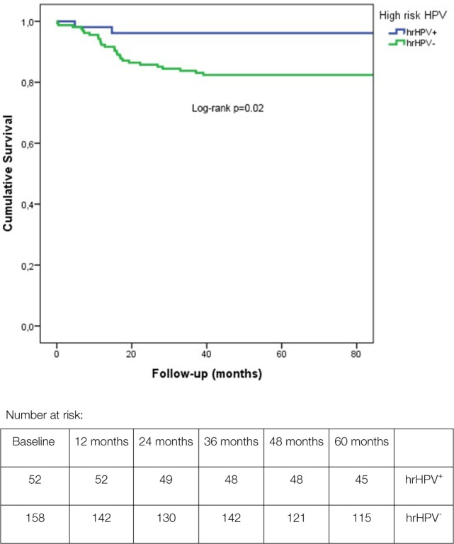
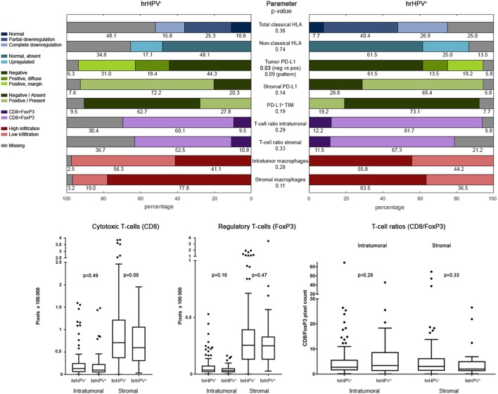
The host's immune system plays a pivotal role in many tumor types, including squamous cell carcinomas (SCCs). We aim to identify immunological prognosticators for lymph node metastases (LNM) and disease-specific survival (DSS) in penile SCC. For this retrospective observational cohort study, penile SCC patients (n = 213) treated in the Netherlands Cancer Institute, were selected if sufficient formalin-fixed, paraffin-embedded tumor material was available. Analysis included previously described high-risk human papilloma virus (hrHPV) status, immunohistochemical scores for classical and non-classical human leukocyte antigen (HLA) class I, programmed death ligand-1 (PD-L1) expression, and novel data on tumor-infiltrating macrophages and cytotoxic an regulatory T-cells. Clinicopathological characteristics and extended follow-up were also included. Regression analyses investigated relationships of the immune parameters with LNM and DSS. In the total cohort, diffuse PD-L1 tumor-cell expression, CD163+ macrophage infiltration, non-classical HLA class I upregulation, and low stromal CD8+ T-cell infiltration were all associated with LNM. In the multivariable model, only tumor PD-L1 expression remained a significant predictor for LNM (odds ratio (OR) 2.8, p = 0.05). hrHPV negativity and diffuse PD-L1 tumor-cell expression were significantly associated with poor DSS and remained so upon correction for clinical parameters [hazard ratio (HR) 9.7, p < 0.01 and HR 2.8, p = 0.03]. The only immune factor with different expression in HPV+ and HPV- tumors was PD-L1, with higher PD-L1 expression in the latter (p = 0.03). In the HPV- cohort (n = 158), LNM were associated with diffuse PD-L1 tumor-cell expression, high intratumoral CD163+ macrophage infiltration, and low number of stromal CD8+ T-cells. The first two parameters were also linked to DSS. In the multivariable regression model, diffuse PD-L1 expression remained significantly unfavorable for DSS (HR 5.0, p < 0.01). These results emphasize the complexity of the tumor microenvironment in penile cancer and point toward several possible immunotherapy targets. Here described immune factors can aid risk-stratification and should be evaluated in clinical immunotherapy studies to ultimately lead to patient tailored treatment.
Keywords: B7-H1; HPV; T-cells; immune escape; microenvironment; penile cancer; programmed death ligand-1; squamous cell carcinoma.
Figures
Similar articles
-
Immune-checkpoint status in penile squamous cell carcinoma: a North American cohort.Hum Pathol. 2017 Jan;59:55-61. doi: 10.1016/j.humpath.2016.09.003. Epub 2016 Sep 20. Hum Pathol. 2017. PMID: 27663086
-
High tumour mutational burden is associated with strong PD-L1 expression, HPV negativity, and worse survival in penile squamous cell carcinoma: an analysis of 165 cases.Pathology. 2024 Apr;56(3):357-366. doi: 10.1016/j.pathol.2023.10.010. Epub 2023 Dec 1. Pathology. 2024. PMID: 38161143
-
The prognostic impact of programmed cell death ligand 1 and human leukocyte antigen class I in pancreatic cancer.Cancer Med. 2017 Jul;6(7):1614-1626. doi: 10.1002/cam4.1087. Epub 2017 Jun 10. Cancer Med. 2017. PMID: 28602029 Free PMC article.
-
The prognostic role of PD-L1 expression for survival in head and neck squamous cell carcinoma: A systematic review and meta-analysis.Oral Oncol. 2018 Nov;86:81-90. doi: 10.1016/j.oraloncology.2018.09.016. Epub 2018 Sep 17. Oral Oncol. 2018. PMID: 30409325
-
A Systematic Review of the Tumor-Infiltrating CD8+ T-Cells/PD-L1 Axis in High-Grade Glial Tumors: Toward Personalized Immuno-Oncology.Front Immunol. 2021 Sep 17;12:734956. doi: 10.3389/fimmu.2021.734956. eCollection 2021. Front Immunol. 2021. PMID: 34603316 Free PMC article.
Cited by
-
Safety and efficacy of immune checkpoint inhibitors in advanced penile cancer: report from the Global Society of Rare Genitourinary Tumors.J Natl Cancer Inst. 2023 Dec 6;115(12):1605-1615. doi: 10.1093/jnci/djad155. J Natl Cancer Inst. 2023. PMID: 37563779 Free PMC article.
-
Immunotherapy in the Management of Penile Cancer-A Systematic Review.Cancers (Basel). 2025 Mar 4;17(5):883. doi: 10.3390/cancers17050883. Cancers (Basel). 2025. PMID: 40075730 Free PMC article. Review.
-
Emerging Advances in the Molecular Landscape of Penile Cancer and Their Implications for Precision Medicine.Curr Treat Options Oncol. 2025 May;26(5):367-374. doi: 10.1007/s11864-025-01319-3. Epub 2025 Apr 16. Curr Treat Options Oncol. 2025. PMID: 40237885 Review.
-
Expansion of tumor-infiltrating lymphocytes (TIL) from penile cancer patients.Int Immunopharmacol. 2021 May;94:107481. doi: 10.1016/j.intimp.2021.107481. Epub 2021 Feb 23. Int Immunopharmacol. 2021. PMID: 33636562 Free PMC article.
-
Machine learning-based overall and cancer-specific survival prediction of M0 penile squamous cell carcinoma:A population-based retrospective study.Heliyon. 2023 Dec 8;10(1):e23442. doi: 10.1016/j.heliyon.2023.e23442. eCollection 2024 Jan 15. Heliyon. 2023. PMID: 38163093 Free PMC article.
References
-
- Hakenberg OW, Compérat E, Minhas S, Necchi A, Protzel C, Watkin NA. EAU Guidelines on Penile Cancer. (2018). Available from: http://uroweb.org/guideline/penile-cancer/ (Accessed: March 25, 2018). - PubMed
MeSH terms
Substances
LinkOut - more resources
Full Text Sources
Other Literature Sources
Research Materials