. 2018 Jun 11:9:1312.
doi: 10.3389/fimmu.2018.01312. eCollection 2018.

DN2 Thymocytes Activate a Specific Robust DNA Damage Response to Ionizing Radiation-Induced DNA Double-Strand Breaks

Affiliations

DN2 Thymocytes Activate a Specific Robust DNA Damage Response to Ionizing Radiation-Induced DNA Double-Strand Breaks

Irene Calvo-Asensio et al. Front Immunol. .

Abstract

For successful bone marrow transplantation (BMT), a preconditioning regime involving chemo and radiotherapy is used that results in DNA damage to both hematopoietic and stromal elements. Following radiation exposure, it is well recognized that a single wave of host-derived thymocytes reconstitutes the irradiated thymus, with donor-derived thymocytes appearing about 7 days post BMT. Our previous studies have demonstrated that, in the presence of donor hematopoietic cells lacking T lineage potential, these host-derived thymocytes are able to generate a polyclonal cohort of functionally mature peripheral T cells numerically comprising ~25% of the peripheral T cell pool of euthymic mice. Importantly, we demonstrated that radioresistant CD44+ CD25+ CD117+ DN2 progenitors were responsible for this thymic auto-reconstitution. Until recently, the mechanisms underlying the radioresistance of DN2 progenitors were unknown. Herein, we have used the in vitro "Plastic Thymus" culture system to perform a detailed investigation of the mechanisms responsible for the high radioresistance of DN2 cells compared with radiosensitive hematopoietic stem cells. Our results indicate that several aspects of DN2 biology, such as (i) rapid DNA damage response (DDR) activation in response to ionizing radiation-induced DNA damage, (ii) efficient repair of DNA double-strand breaks, and (iii) induction of a protective G1/S checkpoint contribute to promoting DN2 cell survival post-irradiation. We have previously shown that hypoxia increases the radioresistance of bone marrow stromal cells in vitro, at least in part by enhancing their DNA double-strand break (DNA DSB) repair capacity. Since the thymus is also a hypoxic environment, we investigated the potential effects of hypoxia on the DDR of DN2 thymocytes. Finally, we demonstrate for the first time that de novo DN2 thymocytes are able to rapidly repair DNA DSBs following thymic irradiation in vivo.

Keywords: DN2 pro-T cells; DNA damage response; bone marrow transplantation; hypoxia; ionizing radiation; thymic auto-reconstitution.


Figures

Figure 1
Figure 1
Double-negative (DN)2 thymocytes survive γ-irradiation in vitro. Clonogenic survival assays of mouse DN2 thymocytes and NH-hematopoietic stem cells (HSCs) γ-irradiated at 0.5–4 Gy and cultured for 3 or 5 days (depending on the cell type). Error bars represent mean ± SD, n = 3. *p < 0.05, two-way ANOVA analysis with Sidak’s multiple comparisons test.
Figure 2
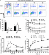
Figure 2
Double-negative (DN)2 thymocytes activate DNA damage checkpoints. (A) Representative cytograms of DN2 and NH-hematopoietic stem cells (HSCs) at 0–24 h post 4 Gy irradiated and stained for bromo-deoxyuridine (BrdU) incorporation and DNA content using propidium iodide (PI). Colored boxes are used to indicate G1, S, and G2/M phase cells under control conditions; black arrowheads indicate sub-G1 cells and * indicates cohort of BrdU-labeled DN2 cells accumulated in late S/G2. (B) Quantification of the percentage DN2 and NH-HSCs in each phase of the cell cycle under normal conditions. (C) Quantification of the percentage G1 DN2 cells and NH-HSCs at 0–36 h post-irradiation or under normal conditions. (D) Quantification of the percentage BrdU-labeled G1/early S phase DN2 cells and NH-HSCs at 0–36 h post-irradiation or under normal conditions. (E) Quantification of the percentage sub-G1 DN2 cells and NH-HSCs at 0–36 h post-irradiation or under normal conditions All cytograms and graphs are representative of three independent experiments. Error bars represent mean ± SD, n = 3, *p < 0.05, **p < 0.01, ****p < 0.0001. Two-way ANOVA analysis with Tukey’s multiple comparisons test was performed on the following conditions (i) DN2-ionizing radiation (IR) versus DN2 + 4 Gy and (ii) HSC-IR versus HSC + 4 Gy.
Figure 3
Figure 3
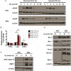
DN2 thymocytes activate a robust DNA damage response following γ-irradiation. Western blot analysis of (A) H2AX (Ser139) phosphorylation (γH2AX—marker of DNA double-strand breaks), p53 stabilization; and p21 and Puma expression in DN2 cells and NH-hematopoietic stem cells (HSCs) at 0–24 h post 4 Gy irradiation. Western blot analysis of total endogenous levels of (B) of H2AX, ATM, DNA-PKcs and Chk2; (C) of 53BP1, DNA ligase IV and Rad51; and (D) of Bcl-2, Bcl-XL, and Bim in un-irradiated (control) DN2 cells and NH-HSCs. β-Actin and β-tubulin were used as internal controls. All images are representative of one of three independent experiments.
Figure 4
Figure 4
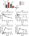
Cell cycle checkpoint activation of normoxic and hypoxic DN2 thymocytes. (A) Comparison of the percentage of cells in each phase of the cell cycle in NH-hematopoietic stem cell (HSC) and DN2 cells cultured in 21 or 5% O2. *p < 0.05, multiple t-tests with Holm–Sidak post-test correction. Quantification of average percentage of G1 phase (B) DN2 and (C) NH-HSC cells cultured in either 21 or 5% O2, 0–36 h post bromo-deoxyuridine (BrdU) pulse, with or without treatment with 4 Gy of ionizing radiation (IR). *p < 0.05, **p < 0.01, ***p < 0.001, two-way ANOVA analysis with Bonferroni post-test correction, n = 3. Quantification of average percentage of BrdU-labeled G1 phase (D) DN2 and (E) NH-HSC cells cultured in either 21 or 5% O2, 0–36 h post BrdU pulse, with or without treatment with 4 Gy of IR. **p < 0.01, ***p < 0.001, two-way ANOVA analysis with Bonferroni post-test correction, n = 3.
Figure 5
Figure 5
DNA repair and apoptotic proteins expression in normoxic and hypoxic NH-hematopoietic stem cell (HSC) and DN2 thymocytes. (A) Western blot analysis of H2AX (Ser139) phosphorylation in DN2 cells and NH-HSCs cultured in either normoxia (21% O2) or hypoxia (5% O2) at 0–24 h post 4 Gy irradiation. (B) mRNA expression levels of DNA repair factors DNA-PKcs (Prkdc), DNA Ligase IV (Lig4) and Rad51 in NH-HSC and DN2 cells in normoxia (21% O2) and hypoxia (5% O2). All values were normalized against β-actin and expressed relative to the NH-HSC normoxic sample. Representative western blots of (C) DNA damage response factors DNA ligase IV and 53BP1; and (D) pro- and anti-apoptotic proteins in NH-HSC and DN2 cells in normoxia (21% O2) and hypoxia (5% O2). All graphs show the average of three biological replicates. (*p < 0.05, multiple t-tests with Holm–Sidak post-test correction).
Figure 6
Figure 6
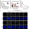
Characterization of DN pro-T cells in vivo response to ionizing radiation (IR). (A) Number of DN1, DN2, and DN3 pro-T cells; (B) number of DN1 and DN2 pro-T cells only that were recovered at different time points 0–12 h after irradiation with 9 Gy. (C) Average number of γH2AX IR-induced foci (IRIF) per nucleus and (D) representative images of DN2 nuclei stained for γH2AX IRIF and DAPI, corresponding to cells irradiated in vivo and isolated 0–12 h post IR.

References

    1. Rieger MA, Schroeder T. Hematopoiesis. Cold Spring Harb Perspect Biol (2012) 4:1–18.10.1101/cshperspect.a008250 - DOI - PMC - PubMed
    1. Jagannathan-Bogdan M, Zon LI. Hematopoiesis. Development (2013) 140:2463–7.10.1242/dev.083147 - DOI - PMC - PubMed
    1. Chi AW, Bell JJ, Zlotoff DA, Bhandoola A. Untangling the T branch of the hematopoiesis tree. Curr Opin Immunol (2009) 21:121–6.10.1016/j.coi.2009.01.012 - DOI - PMC - PubMed
    1. Zhao M, Li LH. Regulation of hematopoietic stem cells in the niche. Sci China Life Sci (2015) 58:1209–15.10.1007/s11427-015-4960-y - DOI - PubMed
    1. Miller JFAP. Immunological function of the thymus. Lancet (1961) 2:748–9.10.1016/S0140-6736(61)90693-6 - DOI - PubMed

LinkOut - more resources