. 2018 Jun 11:9:1322.
doi: 10.3389/fimmu.2018.01322. eCollection 2018.

Ectonucleotidase-Mediated Suppression of Lupus Autoimmunity and Vascular Dysfunction

Affiliations

Ectonucleotidase-Mediated Suppression of Lupus Autoimmunity and Vascular Dysfunction

Jason S Knight et al. Front Immunol. .

Abstract

Objectives: CD39 and CD73 are surface enzymes that jut into the extracellular space where they mediate the step-wise phosphohydrolysis of the autocrine and paracrine danger signals ATP and ADP into anti-inflammatory adenosine. Given the role of vascular and immune cells' "purinergic halo" in maintaining homeostasis, we hypothesized that the ectonucleotidases CD39 and CD73 might play a protective role in lupus.

Methods: Lupus was modeled by intraperitoneal administration of pristane to three groups of mice: wild-type (WT), CD39-/-, and CD73-/-. After 36 weeks, autoantibodies, endothelial function, kidney disease, splenocyte activation/polarization, and neutrophil activation were characterized.

Results: As compared with WT mice, CD39-/- mice developed exaggerated splenomegaly in response to pristane, while both groups of ectonucleotidase-deficient mice demonstrated heightened anti-ribonucleoprotein production. The administration of pristane to WT mice triggered only subtle dysfunction of the arterial endothelium; however, both CD39-/- and CD73-/- mice demonstrated striking endothelial dysfunction following induction of lupus, which could be reversed by superoxide dismutase. Activated B cells and plasma cells were expanded in CD73-/- mice, while deficiency of either ectonucleotidase led to expansion of TH17 cells. CD39-/- and CD73-/- mice demonstrated exaggerated neutrophil extracellular trap release, while CD73-/- mice additionally had higher levels of plasma cell-free DNA.

Conclusion: These data are the first to link ectonucleotidases with lupus autoimmunity and vascular disease. New therapeutic strategies may harness purinergic nucleotide dissipation or signaling to limit the damage inflicted upon organs and blood vessels by lupus.

Keywords: CD39; CD73; TH17 cells; ectonucleotidases; endothelial dysfunction; neutrophil extracellular traps; systemic lupus erythematosus.


Figures

Figure 1
Figure 1
Pulmonary hemorrhage and splenomegaly in pristane-treated mice. (A) Schematic of CD39 and CD73, which together mediate the step-wise phosphohydrolysis of extracellular ATP into adenosine. (B) Some mice developed clinically overt pulmonary hemorrhage within the first month after pristane administration; these mice required euthanasia. N = 10 saline-treated mice and 20 pristane-treated mice per genotype; no comparisons were statistically significant. (C) Mice were administered either saline or pristane, as indicated. 36 weeks later, spleen size was measured. N = 10 per control group and 17–19 per pristane group; *p < 0.05 and **p < 0.01. Box-and-whisker plots denote minimum, 25th percentile, median, 75th percentile, and maximum.
Figure 2
Figure 2
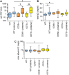
Ectonucleotidase deficiency potentiates autoimmunity in pristane-treated mice. Mice were administered either saline or pristane, as indicated. 36 weeks later, various endpoints were tested. (A) Anti-ribonucleoprotein (Anti-RNP) IgG was measured in serum. (B) Total IgG was measured in serum. (C) Spot albumin/Cr (albumin/creatinine) ratios in urine. N = 10 per control group and 17–19 per pristane group. *p < 0.05 and **p < 0.01.
Figure 3
Figure 3
Modulation of plasma cells and B cells by ectonucleotidase deficiency in pristane-treated mice. Mice were administered either saline or pristane, as indicated. 36 weeks later, splenocytes were analyzed by flow cytometry. (A) CD138+ B220 plasma cells, presented as the percentage of total splenocytes. (B) Representative data as presented in panel (A). (C) CD80+ activated B cells, presented as the percentage of CD19+ B cells. (D) Representative data as presented in panel (C). N = 10 per control group and 17–19 per pristane group; *p < 0.05.
Figure 4
Figure 4
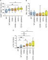
Potentiation of T cell activation and TH17 polarization by ectonucleotidase deficiency. Mice were administered either saline or pristane, as indicated. 36 weeks later, splenocytes were analyzed by flow cytometry. (A) CD44hi CD62Llo effector/memory T cells, presented as the percentage of CD4+ T cells. (B) Percentage of CD4+ T cells expressing interferon gamma. (C) Percentage of CD4+ T cells expressing interleukin 17A. For panel (A), n = 10 per control group and 17–19 per pristane group. For panels (B,C), n = 5 per control group and 8–10 per pristane group; *p < 0.05 and **p < 0.01.
Figure 5
Figure 5
Ectonucleotidase deficiency potentiates endothelial dysfunction in pristane-treated mice. Mice were administered either saline or pristane, as indicated. 36 weeks later, “aortic rings” were harvested for ex vivo determination of endothelial function by measuring the contractile force remaining in pre-contracted (by phenylephrine/PE) aortic rings in response to progressively increasing concentrations of acetylcholine. A “deeper” curve indicates a healthier endothelium, while a “flatter” curve denotes endothelial dysfunction. (A–C) N = 5 control mice and 7 pristane mice per graph. (D,E) Two aortic rings were isolated from each mouse (n = 5), and one was treated with superoxide dismutase (SOD). *p < 0.05, **p < 0.01, and ***p < 0.001.
Figure 6
Figure 6
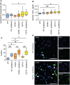
Ectonucleotidase deficiency potentiates neutrophil activation. Mice were administered either saline or pristane, as indicated. 36 weeks later, various endpoints were assessed. (A) Neutrophil-to-lymphocyte ratio in peripheral blood. (B) Cell-free DNA in mouse serum. (C) Mature neutrophils were purified from mouse bone marrow and cultured for 4 h on polylysine-coated glass coverslips. Spontaneous neutrophil extracellular trap (NET) release was assessed by immunofluorescence microscopy. (D) Representative photomicrographs from the data presented in panel C. DNA is stained blue and citrullinated histone H3 (Cit-H3) green. NETs are identified as extracellular areas of blue and green overlap (arrowheads). Scale bar = 50 µm. For panels (A,B), n = 10 per control group and 17–19 per pristane group. For panel (C), n = 6–9 per group; *p < 0.05, **p < 0.01, and ***p < 0.001.

References

    1. Manzi S, Selzer F, Sutton-Tyrrell K, Fitzgerald SG, Rairie JE, Tracy RP, et al. Prevalence and risk factors of carotid plaque in women with systemic lupus erythematosus. Arthritis Rheum (1999) 42:51–60.10.1002/1529-0131(199901)42:1<51::AID-ANR7>3.0.CO;2-D - DOI - PubMed
    1. Reeves WH, Lee PY, Weinstein JS, Satoh M, Lu L. Induction of autoimmunity by pristane and other naturally occurring hydrocarbons. Trends Immunol (2009) 30:455–64.10.1016/j.it.2009.06.003 - DOI - PMC - PubMed
    1. Zhuang H, Szeto C, Han S, Yang L, Reeves WH. Animal models of interferon signature positive lupus. Front Immunol (2015) 6:291.10.3389/fimmu.2015.00291 - DOI - PMC - PubMed
    1. Nacionales DC, Kelly-Scumpia KM, Lee PY, Weinstein JS, Lyons R, Sobel E, et al. Deficiency of the type I interferon receptor protects mice from experimental lupus. Arthritis Rheum (2007) 56:3770–83.10.1002/art.23023 - DOI - PMC - PubMed
    1. Lee PY, Kumagai Y, Li Y, Takeuchi O, Yoshida H, Weinstein J, et al. TLR7-dependent and FcgammaR-independent production of type I interferon in experimental mouse lupus. J Exp Med (2008) 205:2995–3006.10.1084/jem.20080462 - DOI - PMC - PubMed