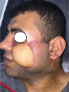
Management of dermatofibrosarcoma protuberans of the face using lower trapezius musculocutaneous pedicle flap reconstruction: a case report
- PMID: 29942467
- PMCID: PMC6007671
- DOI: 10.1093/jscr/rjy089
Management of dermatofibrosarcoma protuberans of the face using lower trapezius musculocutaneous pedicle flap reconstruction: a case report
Abstract
Dermatofibrosarcoma protuberans (DFSP) is a rare neoplasm which represents <0.1% of all tumors but it is considered the most common skin sarcoma. It is a slow-growing tumor that arises from the dermis and invades deeper tissues. The precise origin of DFSP is not well known. It is most frequently seen on the trunk, extremities, and head and neck. The standard treatment of the localized huge DFSP consists of a wide local surgical resection with recommended surgical margins of 2-3 cm. Local recurrence after incomplete excision is common. We present a case of 35-year-old man with enormous bulky mass on the face. Upon histological examination, the diagnosis of DFSP was made, and the patient underwent en bloc wide local excision of the mass followed by the use of Trapezius musculocutaneous pedicle flap reconstruction. On 32 months follow-up, no recurrence has been reported.
Figures


References
-
- Eguzo K, Camazine B, Milner D. Giant dermatofibrosarcoma protuberans of the face and scalp: a case report. Int J Dermatol 2014;53:767–72. - PubMed
-
- McArthur G. Dermatofibrosarcoma protuberans: recent clinical progress. Ann Surg Oncol 2007;14:2876–86. - PubMed
-
- Criscione VD, Weinstock MA. Descriptive epidemiology of dermatofibrosarcoma protuberans in the United States, 1973 to 2002. J Am Acad Dermatol 2007;56:968–73. - PubMed
-
- Burkhardt BR, Soule EH, Winkelmann RK, Ivins JC. Dermatofibrosarcoma protuberans. Study of fifty-six cases. Am J Surg 1966;111:638–44. - PubMed
Publication types
LinkOut - more resources
Full Text Sources
Other Literature Sources
Research Materials

