Review
. 2018 Jun 12:5:13.
doi: 10.1186/s40662-018-0107-0. eCollection 2018.

Optical coherence tomography for ocular surface and corneal diseases: a review

Affiliations
Review

Optical coherence tomography for ocular surface and corneal diseases: a review

Nandini Venkateswaran et al. Eye Vis (Lond). .

Abstract

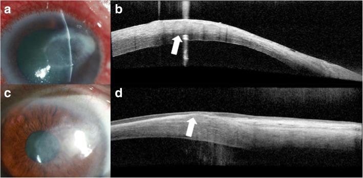
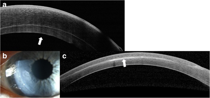
The advent of optical coherence tomography (OCT) imaging has changed the way ophthalmologists image the ocular surface and anterior segment of the eye. Its ability to obtain dynamic, high and ultra-high resolution, cross-sectional images of the ocular surface and anterior segment in a noninvasive and rapid manner allows for ease of use. In this review, we focus on the use of anterior segment OCT, which provides an "optical biopsy" or in vivo imaging of various ocular surface and corneal pathologies, allowing the clinician to diagnose diseases otherwise not visualized by traditional methods. The utility of anterior segment OCT for various anterior segment pathologies is reviewed.

Keywords: Anterior segment optical coherence tomography; Ocular surface imaging; Ocular surface lesions.


Conflict of interest statement

Not applicable.The authors declare they have no competing interests.

Figures

Fig. 1
Fig. 1
AS-OCT of a normal tear film and cornea. AS-OCT displaying a normal tear film and cornea
Fig. 2
Fig. 2
Slit lamp photograph and AS-OCT of keratoconic corneas with corneal scarring. a Slit lamp photograph of central scarring in a cornea affected by keratoconus. b AS-OCT shows an area of anterior corneal scarring and thinning (arrow). c Slit lamp photograph of corneal haze three days after corneal collagen cross linking (arrow). d AS-OCT shows a subtle demarcation line in the area of corneal haze (arrow)
Fig. 3
Fig. 3
Intrastromal corneal ring segments used in keratoconus. a Slit lamp photograph of an intrastromal corneal ring segment used for the treatment of keratoconus. b AS-OCT image captures the corneal intrastromal segment and helps assess its location and depth within the cornea (arrow)
Fig. 4
Fig. 4
Slit lamp photograph and AS-OCT of ocular surface squamous neoplasia pre and post treatment. a Slit lamp photograph of a papillomatous conjunctival lesion. b There is an abrupt transition from normal epithelium with thickened hyperreflective epithelium (arrow) on AS-OCT characteristic of ocular surface squamous neoplasia. c Slit lamp photograph showing complete resolution of the papillomatous conjunctival lesion after two cycles of 5-fluorouracil. d There is normalization of the conjunctival and corneal architecture (arrow) after two cycles of topical 5-fluorouracil on AS-OCT
Fig. 5
Fig. 5
Slit lamp photograph and AS-OCT of conjunctival melanoma. a Slit lamp photograph of a mixed amelanotic/pigmented conjunctival melanoma. b AS-OCT shows a hyperreflective, subepithelial lesion (asterisk) with thin but hyperreflective epithelium (arrow)
Fig. 6
Fig. 6
Slit lamp photograph and AS-OCT of conjunctival lymphoma. a Slit lamp photograph of conjunctival lymphoma. b On AS-OCT, there is a homogeneous, dark, hyporeflective subepithelial lesion with smooth borders and overlying thin epithelium (arrow). The lesion contains monomorphic, stippled, dot-like infiltrates corresponding to the infiltration of monoclonal lymphocytes
Fig. 7
Fig. 7
Slit lamp photograph and AS-OCT of conjunctival amyloidosis. a Slit lamp photograph of conjunctival amyloidosis (arrow). b AS-OCT image of conjunctival amyloidosis showing a heterogeneous, dark subepithelial lesion with irregular borders containing hyper-reflective linear infiltrates that correspond to amyloid deposition (arrow)
Fig. 8
Fig. 8
Slit lamp photograph and AS-OCT of pterygium. a Slit lamp photograph of a pterygium. b AS-OCT image of the pterygium shows a dense, hyper-reflective, fibrillary subepithelial lesion that is between the corneal epithelium and Bowman’s layer (arrow)
Fig. 9
Fig. 9
Slit lamp photograph and AS-OCT of a conjunctival nevus. a Slit lamp photograph displaying a cystic nevus in a child. b On AS-OCT, this lesion is a well-circumscribed subepithelial lesion containing cystic spaces (arrow)
Fig. 10
Fig. 10
Slit lamp photograph and AS-OCT of primary acquired melanosis. a Slit lamp photograph of primary acquired melanosis (arrow). b AS-OCT image shows areas of subepithelial reflectivity (arrow)
Fig. 11
Fig. 11
Slit lamp photograph and AS-OCT of a Salzmann’s nodule and band keratopathy. a Slit lamp photograph of a central Salzmann’s nodule. b On AS-OCT, the nodule is seen as a localized area of hyperreflective material that has replaced the anterior stroma and Bowman’s layer underneath normal epithelium (arrow). c Slit lamp photograph of band keratopathy in the peripheral cornea (arrow). d AS-OCT imaging shows a thin band of hyperreflectivity along Bowman’s layer with underlying shadowing (arrow)
Fig. 12
Fig. 12
Slit lamp photograph and AS-OCT of granular stromal dystrophy. a Slit lamp photograph of granular stromal dystrophy with positive Masson-Trichrome and negative amyloid staining. b On AS-OCT, there is hyperreflective material deposited in the anterior stroma with clear intervening spaces (arrow)
Fig. 13
Fig. 13
Slit lamp photograph and AS-OCT of infectious keratitis and subsequent corneal scarring. a Slit lamp photograph of a patient with contact lens related Pseudomonas infectious keratitis. b AS-OCT shows diffuse stromal hyperreflectivity and thickening in the area of the infiltrate involving nearly 50% of the stroma (arrow). c Slit lamp photograph of a compact, subepithelial scar after infectious keratitis. d AS-OCT shows subepithelial thinning and hyperreflectivity in the area of the corneal scar (arrow)
Fig. 14
Fig. 14
Slit lamp photograph and AS-OCT of an attached DSAEK graft as well as epithelial ingrowth. a AS-OCT of an attached DSAEK graft (arrow) post-operatively. b Slit lamp photograph of epithelial ingrowth after LASIK. c AS-OCT demonstrating the area epithelial ingrowth after LASIK (arrow)

References

    1. Karp CL. Evolving Technologies for lid and Ocular Surface Neoplasias: is optical biopsy a reality? JAMA ophthalmology. 2017;135(8):852–853. doi: 10.1001/jamaophthalmol.2017.2009. - DOI - PubMed
    1. Izatt JA, Hee MR, Swanson EA, Lin CP, Huang D, Schuman JS, et al. Micrometer-scale resolution imaging of the anterior eye in vivo with optical coherence tomography. Arch Ophthalmol. 1994;112(12):1584–1589. doi: 10.1001/archopht.1994.01090240090031. - DOI - PubMed
    1. Han SB, Liu YC, Noriega KM, Mehta JS. Applications of anterior segment optical coherence tomography in cornea and ocular surface diseases. J Ophthalmol. 2016;2016:4971572. - PMC - PubMed
    1. Wang J, Abou Shousha M, Perez VL, Karp CL, Yoo SH, Shen M, et al. Ultra-high resolution optical coherence tomography for imaging the anterior segment of the eye. Ophthalmic Surg Lasers Imaging. 2011;42(Suppl):S15–S27. doi: 10.3928/15428877-20110627-02. - DOI - PubMed
    1. Kanellopoulos AJ, Asimellis G. Anterior-segment optical coherence tomography investigation of corneal deturgescence and epithelial remodeling after DSAEK. Cornea. 2014;33(4):340–348. doi: 10.1097/ICO.0000000000000053. - DOI - PubMed

LinkOut - more resources