. 2018 Oct;79(4):197-207.
doi: 10.1111/cod.13046. Epub 2018 Jun 25.

Regulation of contact sensitivity in non-obese diabetic (NOD) mice by innate immunity

Affiliations

Regulation of contact sensitivity in non-obese diabetic (NOD) mice by innate immunity

Marian Szczepanik et al. Contact Dermatitis. 2018 Oct.

Abstract

Background: Genetic background influences allergic immune responses to environmental stimuli. Non-obese diabetic (NOD) mice are highly susceptible to environmental stimuli. Little is known about the interaction of autoimmune genetic factors with innate immunity in allergies, especially skin hypersensitivity.

Objectives: To study the interplay of innate immunity and autoimmune genetic factors in contact hypersensitivity (CHS) by using various innate immunity-deficient NOD mice.

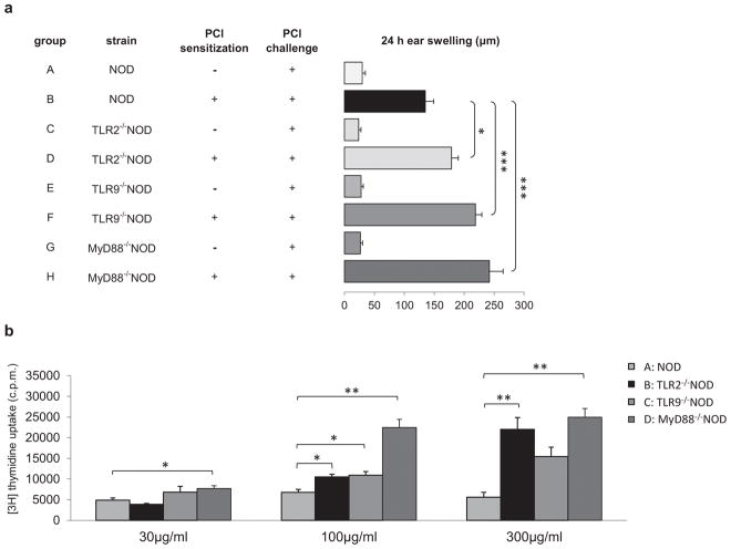
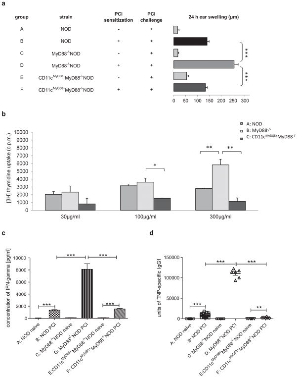
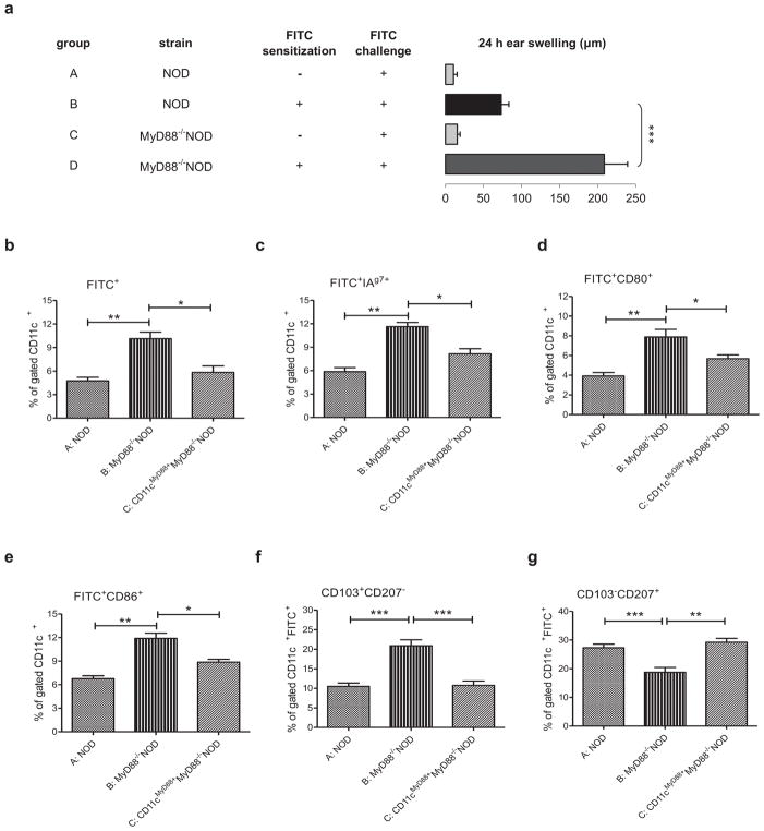
Methods: Toll-like receptor (TLR) 2-deficient, TLR9-deficient and MyD88-deficient NOD mice were used to investigate CHS. The cellular mechanism was determined by flow cytometry in vitro and adoptive cell transfer in vivo. To investigate the role of MyD88 in dendritic cells (DCs) in CHS, we also used CD11cMyD88+ MyD88-/- NOD mice, in which MyD88 is expressed only in CD11c+ cells.

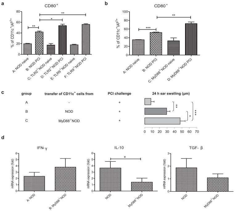
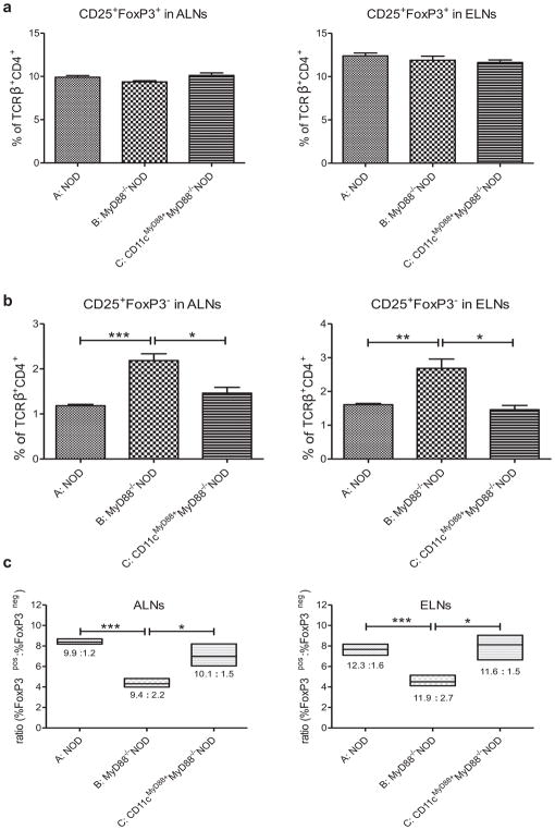
Results: We found that innate immunity negatively regulates CHS, as innate immunity-deficient NOD mice developed exacerbated CHS accompanied by increased numbers of skin-migrating CD11c+ DCs expressing higher levels of major histocompatibility complex II and CD80. Moreover, MyD88-/- NOD mice had increased numbers of CD11c+ CD207- CD103+ DCs and activated T effector cells in the skin-draining lymph nodes. Strikingly, re-expression of MyD88 in CD11c+ DCs (CD11cMyD88+ MyD88-/- NOD mice) restored hyper-CHS to a normal level in MyD88-/- NOD mice.

Conclusion: Our results suggest that the autoimmune-prone NOD genetic background aggravates CHS regulated by innate immunity, through DCs and T effector cells.

Keywords: Toll-like receptor; Tregs; contact sensitivity; dendritic cells; non-obese diabetic (NOD) mouse.


Conflict of interest statement

CONFLICT OF INTEREST

The authors declare that they have no conflicts of interest.

Figures

Fig. 1
Fig. 1
Role of TLRs and MyD88 in CHS in NOD mice. (A) TLR2 and TLR9 and MyD88 deficiency in NOD mice showed an increased CHS response to PCl. WT NOD (group B) or TLR2−/− (group D) or TLR9−/− (group F) or MyD88−/− (group H) NOD mice were sensitized with 5% PCl. Four days later all the mice were challenged with 0.4% PCl and tested for CHS response 24 hours later. N = 8–12, *P <.05 and ***P <.001 (ANOVA followed by Tukey’s test). (B) TLR2−/− and TLR9−/− and MyD88−/− NOD mice develop stronger antigen specific responses in vitro. WT NOD mice (group A) or TLR2−/− (group B) or TLR9−/− (group C) or MyD88−/− (group D) NOD mice were sensitized with 5% PCl. Four days later ALNs were isolated and ALNC were cultured in the presence of the antigen, TNP40-Ig, at different concentration for 48 hours. The mean background in cultures in the absence of the TNP-Ig for NOD, TLR2−/−NOD, TLR9−/−NOD and MyD88−/−NOD was 10744±257, 24997±774, 10387±600 and 6440±570 respectively. N = 3, *P <.05 and **P <.01 (Two-tailed Student’s t-test). All error bars represent SE.
Fig. 2
Fig. 2
Role of DCs in CHS in TLR- or MyD88-deficient NOD mice. (A) Increased percentage of CD11c+I-Ag7+ DCs expressing CD80 in TLR2 and TLR9 deficient NOD mice. WT NOD (group B) or TLR2−/− (group D) or TLR9−/− (group F) NOD mice were sensitized with 5% PCl. Mice in negative controls (groups A, C and E) were not treated with PCl. ALNs were isolated 3 days later and ALNC were stained with different monoclonal antibodies (anti-CD11c-APC, anti-CD80-PE, anti-I-Ag7-FITC). Gating information for the flow analysis is presented in Fig. S2. N=3, *P <.05 and **P <.01 (Two-tailed Student’s t-test). (B) Increased percentage of CD11c+ I-Ag7+ DCs expressing CD80 in MyD88−/−NOD mice. WT NOD (group B) or MyD88−/−NOD (group D) mice were sensitized with 5% PCl. Mice in negative controls (groups A and C) were not treated with PCl. ALNs were isolated 3 days later and ALNC were stained with different monoclonal antibodies (anti-CD11c-APC, anti-CD80-PE, anti-I-Ag7-FITC). Gating information for the flow analysis is presented in Fig. S2. N=3, **P <.01 and ***P <.001 (Two-tailed Student’s t-test). (C) CD11c+ cells isolated from MyD88−/−NOD mice are stronger inducers of CHS response than CD11c+ cells isolated from WT NOD mice. Donor mice were immunized with PCl three days before the isolation of CD11c+ cells from ALN. Purified CD11c+ cells isolated from NOD (group B) or MyD88−/−NOD (group C) mice were transferred s.c. into naïve NOD recipients, respectively. The recipients were ear-challenged with 0.4% PCl after receiving the cells and tested for CHS responses 24 hours later. N=6–10, *P <.05 and **P <.01 and ***P <.001 (Two-tailed Student’s t-test). (D) Cytokine mRNA expression in skin homogenates. NOD (group A) and MyD88−/−NOD (group B) mice were sensitized with PCl. Five days later mRNA expression was measured in skin homogenates. N=10–12, *P <.05 (Mann Whitney’s test). All error bars represent SE.
Fig. 3
Fig. 3
Re-expression of MyD88 in CD11c+ DCs restores normal CHS responses in MyD88−/−NOD mice. (A) Presence of MyD88 in CD11c+ DCs restores normal CHS response in vivo. WT NOD (group B) or MyD88−/− (group D) or CD11cMyD88+MyD88−/− (group F) NOD mice were sensitized with 5% PCl or not (group A, C and E, respectively). Four days later all the mice were challenged with 0.4% PCl and tested for CHS 24 hours after challenge. N = 6–14, ***P <.001 (Two-tailed Student’s t-test). (B) CD11cMyD88+MyD88−/−NOD mice develop normal hapten specific response in vitro. WT NOD (group A) or MyD88−/− (group B) or CD11cMyD88+MyD88−/− (group C) NOD mice were sensitized with 5% PCl. ALNs were isolated 4 days later and proliferation assay was performed as described in Fig. 2B. N = 3, *P <.05 and **P <.01 (Two-tailed Student’s t-test). (C) Presence of MyD88 in CD11c+ DCs restores normal IFN-γ production. WT NOD (group B) or MyD88−/− (group D) or CD11cMyD88+MyD88−/− (group F) NOD mice were sensitized with 5% PCl or not (group A, C and E, respectively). ALNs were isolated 4 days later and cultured in the presence of antigen, TNP40-Ig for 48 hours. Culture supernatants were harvested and tested for IFN-γ production by ELISA kit. N = 6–12, ***P <.001 (Two-tailed Student’s t-test or Mann Whitney’s test). (D) Presence of MyD88 in CD11c+ DCs restores normal level of TNP-specific IgG1 in serum. WT NOD (group B) or MyD88−/− (group D) or CD11cMyD88+MyD88−/− (group F) NOD mice were sensitized with 5% PCl or not (group A, C and E, respectively). Four days later, all the mice were challenged with 0.4% PCl. The serum was collected 24 hours after challenge. Anti-TNP specific IgG1 antibody was measured by ELISA. N = 6–24. ***P <.001 (Two-tailed Student’s t-test or Mann Whitney’s test). All error bars represent SE.
Fig. 4
Fig. 4
Characterization and tracking of skin DCs in MyD88−/− NOD mice. (A) MyD88-deficient mice on the NOD background develop exaggerated CHS response to FITC. WT NOD and MyD88−/−NOD mice were sensitized with 0.5% FITC (groups B and D) or not (groups A and C). The animals were challenged, 5days later, with FITC and tested for CHS response. N = 4–10, ***P <.001 (ANOVA followed by Tukey’s test). (B–G) Stronger migration and more activated phenotype of skin CD11c+ DCs in MyD88 deficient NOD mice. WT NOD mice (group A), MyD88−/− (group B) and CD11cMyD88+MyD88−/− (group C) NOD mice were sensitized with 0.5% FITC. Three days later, ALNs were isolated and stained with appropriate monoclonal antibodies: anti-I-Ag7-biotin-strep-PE, anti-CD11c-PerCP-Cy5.5, anti-CD11c-APC, anti-CD80-APC, anti-CD86-APC, anti-CD103-PerCP-Cy5.5, anti-CD207-PE. Gating information for the flow analysis is presented in Fig. S2. N = 3, *P <.05 and **P <.01 and ***P <.001 (Two-tailed Student’s t-test). All error bars represent SE.
Fig. 5
Fig. 5
Decreased ratio of CD4+CD25+FoxP3+ Tregs/CD4+CD25+ activated T cells in ALNs and ELNs in MyD88−/−NOD mice. (A) Frequencies of Tregs. (B) Frequencies of T effector cells. (C) Ratio of CD4+CD25+FoxP3+ Tregs/CD4+CD25+ activated T cells. WT NOD mice (group A), MyD88−/− (group B) and CD11cMyD88+MyD88−/− (group C) NOD mice were sensitized with 5% PCl. Four days later, mice were challenged on both sides of the ears with PCl. The ALNs and ELNs were collected 24 hours after challenge. ALNC and ELNC were stained with monoclonal antibodies: anti-TCRβ-FITC, anti-CD4-PerCP-Cy5.5, anti-CD25-APC and anti-FoxP3-PE. Gating information for the flow analysis is presented in Fig. S2. N = 3–4, *P <.05; **P <.01; *** P <.001 (Two-tailed Student’s t-test). All error bars represent SE.

References

    1. Majewska-Szczepanik M, Strzepa A, Marcińska K, Wen L, Szczepanik M. Epicutaneous immunization with TNP-Ig and zymosan induces TCRαβ+ CD4+ contrasuppressor cells that reverse skin-induced suppression via IL-17A. Int Arch Allergy Immunol. 2014;164:122–136. - PMC - PubMed
    1. Kaplan DH, Igyártó BZ, Gaspari AA. Early events in the induction of allergic contact dermatitis. Nat Rev Immunol. 2012;13:114–124. - PMC - PubMed
    1. Askenase PW, Itakura A, Leite-de-Moraes MC, Lisbonne M, Roongapinun S, Goldstein DR, et al. TLR-dependent IL-4 production by invariant Vα14+Jα18+ NKT cells to initiate contact sensitivity in vivo. J Immunol. 2005;175:6390–6401. - PubMed
    1. Jin H, Kumar L, Mathias C, Zurakowski D, Oettgen H, Gorelik L, et al. Toll-like receptor 2 is important for the T(H)1 response to cutaneous sensitization. J Allergy Clin Immunol. 2009;123:875–882. - PMC - PubMed
    1. Klekotka PA, Yang L, Yokoyama WM. Contrasting roles of the IL-1 and IL-18 receptors in MyD88-dependent contact hypersensitivity. J Invest Dermatol. 2010;130:184–91. - PMC - PubMed

MeSH terms