

Appearance of β-dystroglycan precedes the formation of glio-vascular end-feet in developing rat brain
- PMID: 29943956
- PMCID: PMC5966711
- DOI: 10.4081/ejh.2018.2908
Appearance of β-dystroglycan precedes the formation of glio-vascular end-feet in developing rat brain
Abstract
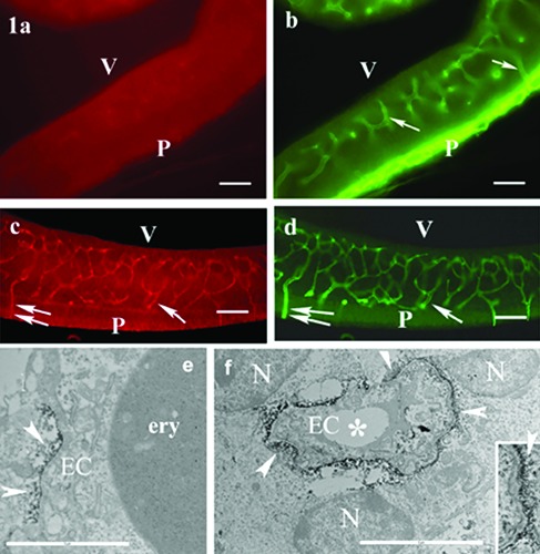
Dystroglycan has an important role in binding of perivascular glial end-feet tothe basal lamina. Its β-subunit is localized in the glial end-feet. The investigation period lasted from E(embryonic day)12 to E20. Laminin and β-dystroglycan were detected by immunohistochemistry, the glial localization of the latter one was supported by electron microscopy. The immatureglial structures were visualized by the immunostaining of nestin. The β-dystroglycan immunoreactivity appeared at E16 following the laminin of basal lamina but preceding the perivascular processes of radial glia (E18) and astrocyte-like cells (E20). It occurred in cell bodies which attached to the vessels directly but not with vascular processes and end-feet. The presence of β-dystroglycan in such immature cells may promote their differentiation to perivascular astrocytes and influence the formation of the glio-vascular processes.
Keywords: Brain development; dystroglycan; glial end-feet; glio-vascular; nestin..
Conflict of interest statement
Conflict of interest: The authors declare that there is no actual or potential conflict of interest.
Figures

Similar articles
-
Glial and perivascular structures in the subfornical organ: distinguishing the shell and core.J Histochem Cytochem. 2015 May;63(5):367-83. doi: 10.1369/0022155415575027. Epub 2015 Feb 11. J Histochem Cytochem. 2015. PMID: 25673286 Free PMC article.
-
Post traumatic lesion absence of beta-dystroglycan-immunopositivity in brain vessels coincides with the glial reaction and the immunoreactivity of vascular laminin.Curr Neurovasc Res. 2008 Aug;5(3):206-13. doi: 10.2174/156720208785425657. Curr Neurovasc Res. 2008. PMID: 18691079
-
Distribution of components of basal lamina and dystrophin-dystroglycan complex in the rat pineal gland: differences from the brain tissue and between the subdivisions of the gland.Histol Histopathol. 2010 Jan;25(1):1-14. doi: 10.14670/HH-25.1. Histol Histopathol. 2010. PMID: 19924636
-
Correlation Between Extravasation and Alterations of Cerebrovascular Laminin and β-Dystroglycan Immunoreactivity Following Cryogenic Lesions in Rats.J Neuropathol Exp Neurol. 2017 Nov 1;76(11):929-941. doi: 10.1093/jnen/nlx081. J Neuropathol Exp Neurol. 2017. PMID: 29044412
-
Laminin and fibronectin in normal and malignant neuroectodermal cells.Med Biol. 1984;62(3):163-80. Med Biol. 1984. PMID: 6387323 Review.
Cited by
-
Ultrastructural histochemistry in biomedical research: Alive and kicking.Eur J Histochem. 2018 Nov 7;62(4):2990. doi: 10.4081/ejh.2018.2990. Eur J Histochem. 2018. PMID: 30418011 Free PMC article.
-
A journal of histochemistry as a forum for non-histochemical scientific societies.Eur J Histochem. 2019 Dec 23;63(4):3106. doi: 10.4081/ejh.2019.3106. Eur J Histochem. 2019. PMID: 31868322 Free PMC article.
-
Histochemistry as a versatile research toolkit in biological research, not only an applied discipline in pathology.Eur J Histochem. 2018 Dec 21;62(4):3006. doi: 10.4081/ejh.2018.3006. Eur J Histochem. 2018. PMID: 30572698 Free PMC article.
-
A large portion of the astrocyte proteome is dedicated to perivascular endfeet, including critical components of the electron transport chain.J Cereb Blood Flow Metab. 2021 Oct;41(10):2546-2560. doi: 10.1177/0271678X211004182. Epub 2021 Apr 4. J Cereb Blood Flow Metab. 2021. PMID: 33818185 Free PMC article.
References
-
- Wolburg H, Noell S, Mack A Wolburg- Buchholz K Fallier-Becker P. Brain endothelial cells and the glio-vascular complex. Cell Tissue Res 2009;335:75-96. - PubMed
-
- Zaccaria ML, Di Tommaso F, Brancaccio A, Paggi P, Petrucci TC. Dystroglycan distribution in adult mouse brain: a light and electron microscopic study. Neuroscience 2001; 104:311-24. - PubMed
-
- Szabó A, Kálmán M. Post traumatic lesion absence of β-dystroglycan immunopositivity in brain vessels coincides with the glial reaction and the immunoreactivity of vascular laminin. Curr Neurovasc Res 2008;5:206-13. - PubMed
-
- Tian M, Jacobson C, Gee SH, Campbell KP, Carbonetto S, Jucker M. Dystroglycan in the cerebellum is a laminin a2-chain binding protein at the glial-vascular interface and is expressed in Purkinje cells. Eur J Neurosci 1996; 8:2739-47. - PubMed
MeSH terms
Substances
LinkOut - more resources
Full Text Sources
Other Literature Sources
