

Review
doi: 10.1148/radiol.2018171820.
Epub 2018 Jun 26.
Current Applications and Future Impact of Machine Learning in Radiology
Affiliations
- PMID: 29944078
- PMCID: PMC6542626
- DOI: 10.1148/radiol.2018171820
Item in Clipboard
Review
Current Applications and Future Impact of Machine Learning in Radiology
Radiology.
2018 Aug.
Abstract
Recent advances and future perspectives of machine learning techniques offer promising applications in medical imaging. Machine learning has the potential to improve different steps of the radiology workflow including order scheduling and triage, clinical decision support systems, detection and interpretation of findings, postprocessing and dose estimation, examination quality control, and radiology reporting. In this article, the authors review examples of current applications of machine learning and artificial intelligence techniques in diagnostic radiology. In addition, the future impact and natural extension of these techniques in radiology practice are discussed.
© RSNA, 2018.
Figures
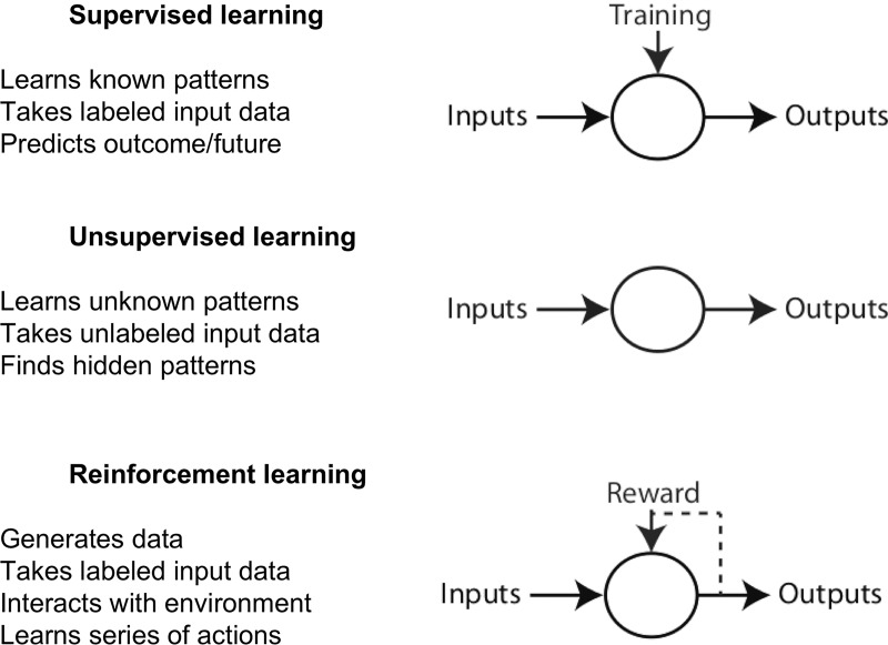
Image shows different categories of machine learning.
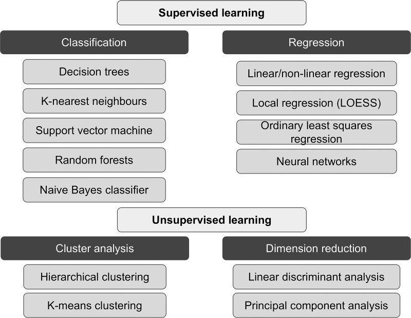
Image shows summary of supervised and unsupervised learning paradigms and subcategories, with examples in each subcategory.
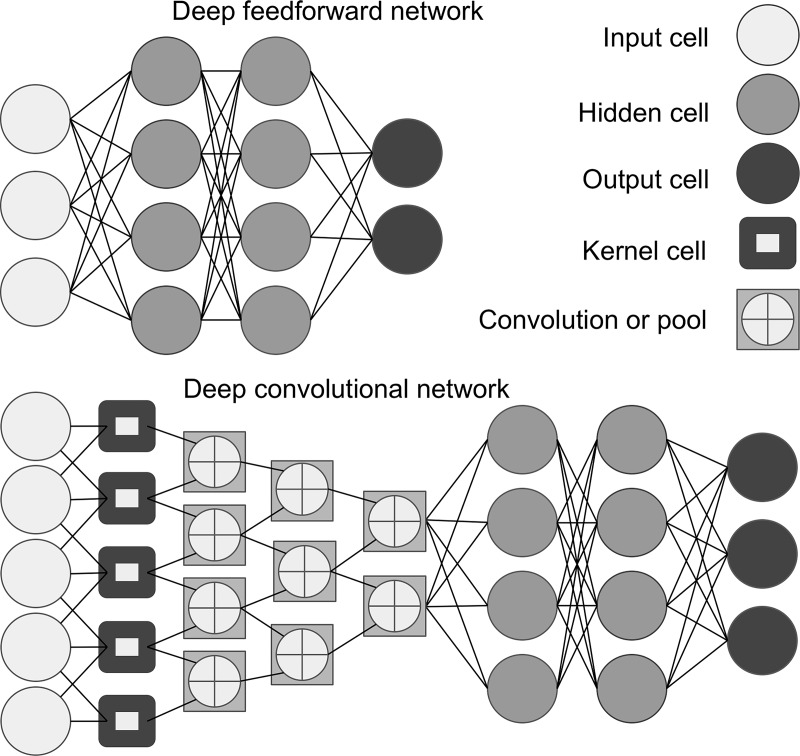
Image shows artificial neural network, an interconnected group of processing elements similar to network of neurons in the brain. Each processing element is called a cell (also called neuron or node). Multiple hidden layers with nodes allow for multiple mathematical calculations to generate outputs. Deep learning is an artificial neural network algorithm that contains more than one hidden layer. Feedforward neural network (top panel) is the simplest type of artificial neural network. In this network, information moves in only one direction (forward) from input nodes, through hidden nodes, and to output nodes. Convolutional neural network (bottom panel) is a type of feedforward artificial neural network built from multiple hidden layers including convolutional layers, pooling layers, fully connected layers, and normalization layers. Convolution layer is comprised of filter elements known as kernels.
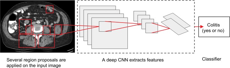
Image shows proposed deep convolutional neural network (CNN) system for detection of colitis. In the first step, several thousand automated regions are applied on each CT section with an algorithm that finds all possible places where objects can be located (region proposal). For each region proposal, feature extraction and computation are performed by implementation of CNN with multiple hidden layers by using pretrained data sets. In the last step, classifier algorithm (eg, linear support vector machine) could be used for colitis classification.
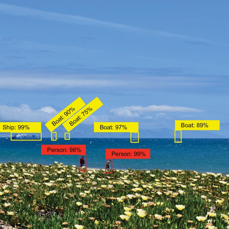
Image shows that feature extraction and object recognition, qualified by using confidence indicators, is made simple with toolkits such as TensorFlow object detection application programming interface. (Image courtesy of Omid Khalilzadeh, MD, MPH, Massachusetts General Hospital, Boston, Mass.)
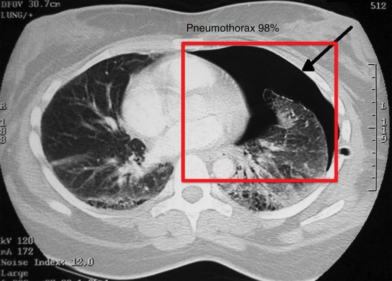
Image shows feature extraction example in medical imaging use case. Automated detection of critical findings such as pneumothorax in medical imaging is one application of machine learning. Heads-up display or method of highlighting relevant findings in a picture archiving and communication system or other image viewing system is example of how machine learning can be productized and integrated into radiology workflow.
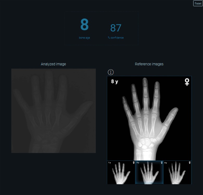
Image shows automated bone age algorithm based on machine learning techniques. Opportunities exist to automate and help replace more manual workflows, such as use of book-based references.
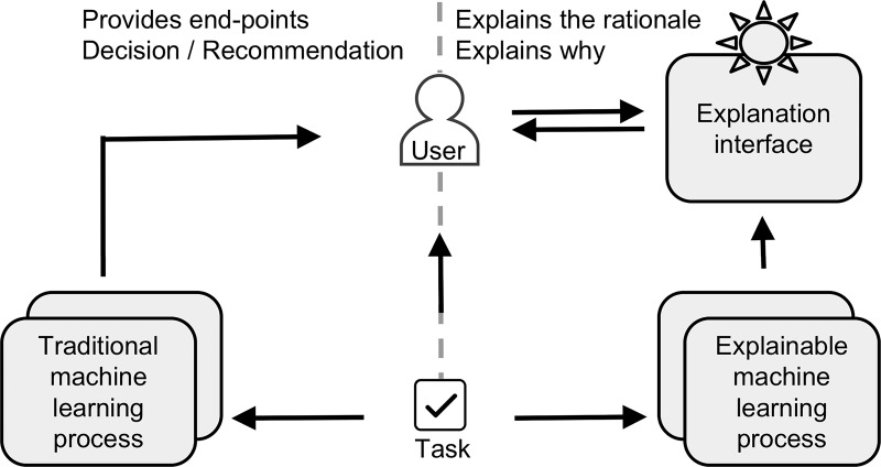
Image compares traditional (left panel) and explainable (right panel) artificial intelligence systems. Defense Advanced Research Projects Agency is developing explainable artificial intelligence systems that will have ability to explain their rationale through explanation interface. This approach will enable human users to more effectively manage and more confidently trust artificial intelligent systems.
References
Publication types
MeSH terms
Grants and funding
LinkOut - more resources
Full Text Sources
Other Literature Sources
