

Gross Morphometry, Histomorphometry, and Immunohistochemistry Confirm Early and Persistent Jejunal Crypt Hyperplasia in Poults with Enteritis and Depressed Growth
- PMID: 29944394
- PMCID: PMC6176685
- DOI: 10.1637/11759-101717-Reg.1
Gross Morphometry, Histomorphometry, and Immunohistochemistry Confirm Early and Persistent Jejunal Crypt Hyperplasia in Poults with Enteritis and Depressed Growth
Abstract
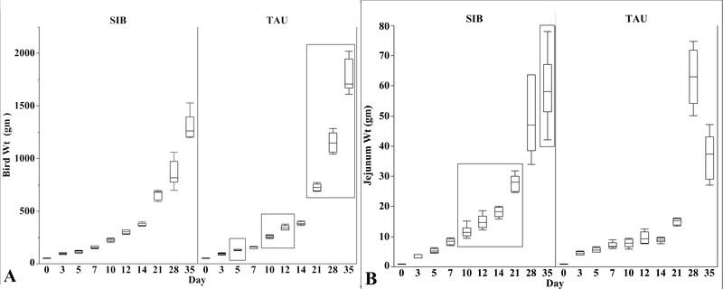
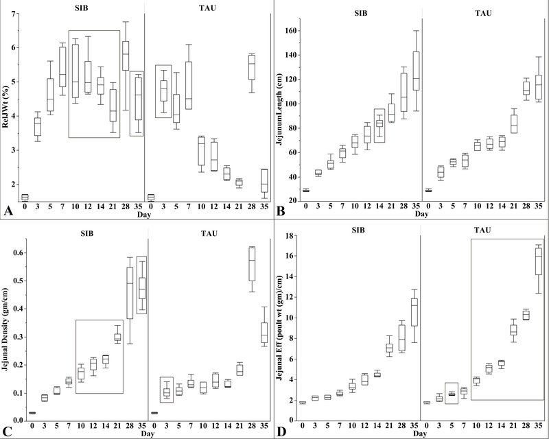
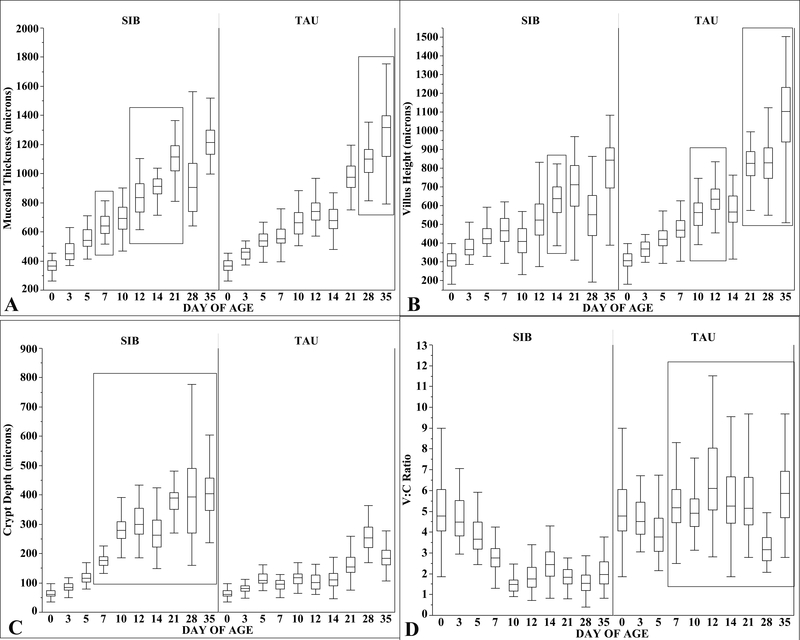
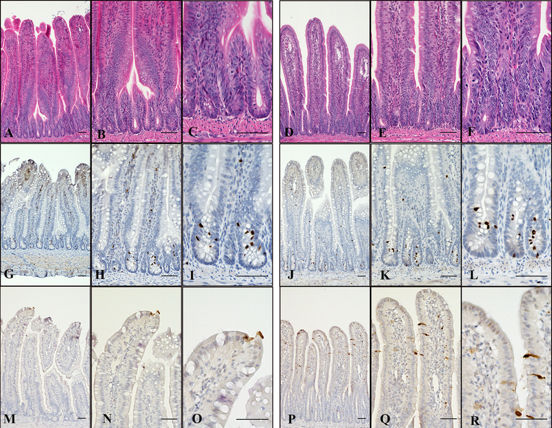
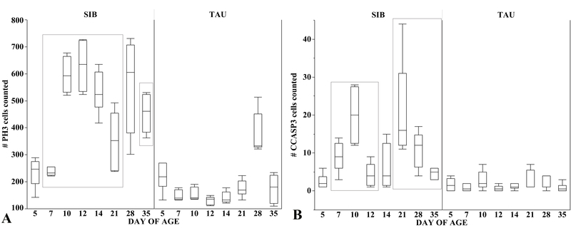
Phosphorylated histone 3 (PH3) and cleaved caspase 3 (CCASP3) were used to detect proliferating and apoptotic cells, respectively, in the jejunums of female sibling poults, with and without enteritis and depressed growth, from hatch to day 35. Poults that developed enteritis and depressed growth (SIB flock) were raised on a commercial farm in eastern North Carolina, whereas poults with normal growth and no enteritis (TAU flock) were raised in the Teaching Animal Unit at North Carolina State University College of Veterinary Medicine. Beginning on day 5 through day 35 and at processing, TAU poults were significantly heavier than SIB poults. Jejunal weights, relative jejunal weights, and jejunal densities were greater in SIB poults from day 10 through 35. Jejunal efficiency (body weight /jejunal length) was higher in TAU poults at day 5 and days 10 through 35. Mucosal thickness was greater in SIB poults between days 7 and 21 but greater in TAU poults at days 28 and 35. From day 7 to 35, villus-to-crypt ratios were higher for TAU poults and lower for SIB poults because hyperplastic crypts formed a greater percentage of the mucosa in SIB poults. By day 7, PH3- and CCASP3-positive cells were increased in SIB poults, showing that mucosal changes resulted from combined crypt epithelial hyperplasia and increased apoptosis of villous enterocytes. Findings in this study confirm that enteritis, in the absence of clinical signs, and depressed growth in turkey poults begins by day 7, can be identified microscopically, persists for at least 35 days, is associated with lower processing weights, and has a profound negative effect on turkey growth.
Keywords: crypt hyperplasia; gross morphometry; growth; gut health; histomorphometry; immunohistochemistry; poult enteritis; turkey.
Figures
Similar articles
-
Poult malabsorption syndrome. I. Malabsorption in poult enteritis.Avian Dis. 1991 Oct-Dec;35(4):685-93. Avian Dis. 1991. PMID: 1786000
-
Experimental reproduction of a spiking mortality syndrome of turkeys.Avian Dis. 1997 Apr-Jun;41(2):269-78. Avian Dis. 1997. PMID: 9201387
-
Enteric disease in specific-pathogen-free turkey poults inoculated with a small round turkey-origin enteric virus.Avian Dis. 1990 Jul-Sep;34(3):683-92. Avian Dis. 1990. PMID: 2173537
-
Bacterial enteritides of poultry.Poult Sci. 1998 Aug;77(8):1159-65. doi: 10.1093/ps/77.8.1159. Poult Sci. 1998. PMID: 9706083 Review.
-
Poult Enteritis and Mortality Syndrome in Turkey Poults: Causes, Diagnosis and Preventive Measures.Animals (Basel). 2021 Jul 10;11(7):2063. doi: 10.3390/ani11072063. Animals (Basel). 2021. PMID: 34359191 Free PMC article. Review.
Cited by
-
16S rRNA Sequencing Analysis of the Gut Microbiota in Broiler Chickens Prophylactically Administered with Antimicrobial Agents.Antibiotics (Basel). 2021 Feb 2;10(2):146. doi: 10.3390/antibiotics10020146. Antibiotics (Basel). 2021. PMID: 33540533 Free PMC article.
References
-
- Adams NR, Ball RA, Annis CL, and Hofstad MS. Ultrastructural changes in the intestines of turkey poults and embryos affected with transmissible enteritis. J. Comp. Pathol. 82:187–192. 1972. - PubMed
-
- Akbar A, and Reynolds DL. Stunting syndrome in turkey poults: isolation and identification of the etiologic agent. Avian Dis. 41:870–881. 1997. - PubMed
-
- Azad S, Mor SK, Naresh J, Devi P, Sobhy NM, Nhungoc Ti L, and Goyal SM. Detection and molecular characterization of astroviruses in turkeys. Arch. Virol. 161:939–946. 2016. - PubMed
-
- Barnes HJ Prevention, control and treatment of poult enteritis-mortality syndrome. Poult. Dig. 56:16–18. 1997.
-
- Barnes HJ PEC: what is it and what is the economic significance? World Poult. 18:14–16. 2002.
Publication types
MeSH terms
Substances
Grants and funding
LinkOut - more resources
Full Text Sources
Other Literature Sources
Research Materials
