

A whole-genome transcriptome analysis of articular chondrocytes in secondary osteoarthritis of the hip
- PMID: 29944724
- PMCID: PMC6019400
- DOI: 10.1371/journal.pone.0199734
A whole-genome transcriptome analysis of articular chondrocytes in secondary osteoarthritis of the hip
Abstract
Objective: To date, exhaustive gene expression analyses of chondrocytes in hip osteoarthritis (OA) have yielded specific gene expression patterns. No study has reported on the exhaustive transcriptome of secondary hip OA based on acetabular dysplasia in a Japanese population, while previous reports have focused on primary or idiopathic hip OA in Caucasian populations. This study aims to search for specific gene expression patterns of secondary hip OA chondrocytes by transcriptome analysis.
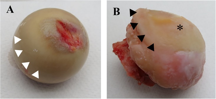
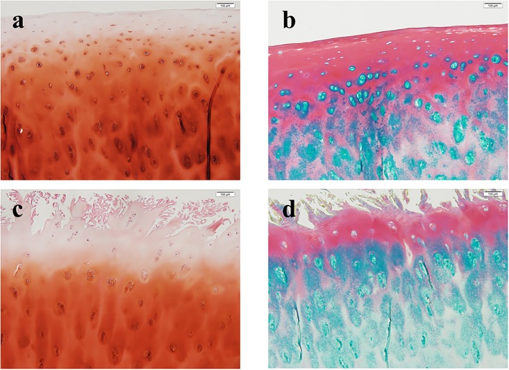
Design: Human articular cartilage was obtained from femoral heads following hemiarthroplasty for femoral neck fracture (N = 8; non-OA) and total hip arthroplasty for secondary hip OA (N = 12). Total RNA was extracted from the articular cartilage and submitted for microarray analysis. The obtained data were used to perform gene expression analysis, GO enrichment analysis and pathway analysis and were compared with data from primary hip OA in Caucasian populations in the literature.
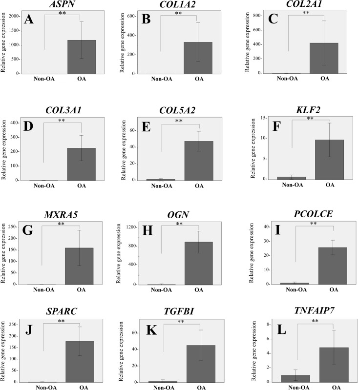
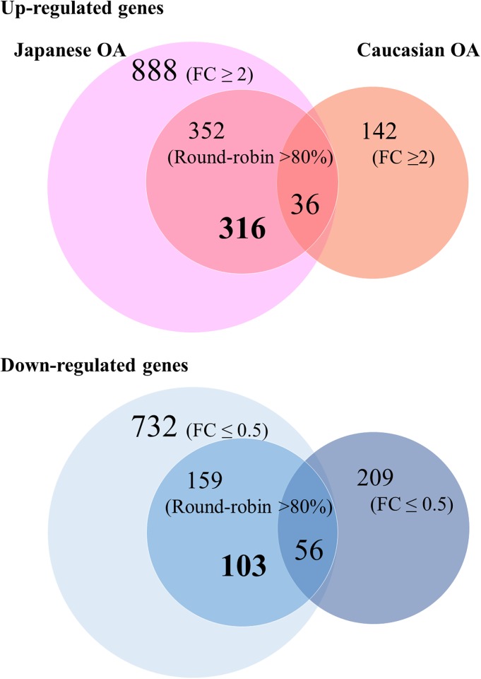
Results: We identified 888 upregulated (fold change: FC ≥ 2) and 732 downregulated (FC ≤ 0.5) genes in hip OA versus non-OA chondrocytes, respectively. Only 10% of upregulated genes were common between the secondary and primary OA. The newly found genes prominently overexpressed in the secondary hip OA chondrocytes were DPT, IGFBP7, and KLF2. Pathway analysis revealed extracellular matrix (ECM)-receptor interaction as an OA-related pathway, which was similar to previous reports in primary hip OA.
Conclusions: This is the first study to report the genome-wide transcriptome of secondary hip OA chondrocytes and demonstrates new potential OA-related genes. Gene expression patterns were different between secondary and primary hip OA, although the results of pathway and functional analysis were similar.
Conflict of interest statement
The authors have declared that no competing interests exist.
Figures
References
-
- Goldring MB, Goldring SR. Osteoarthritis. J Cell Physiol. 2007;213(3):626–634. doi: 10.1002/jcp.21258 - DOI - PubMed
-
- Neogi T, Zhang Y. Epidemiology of osteoarthritis. Rheum Dis Clin North Am. 2013;39(1):1–19. doi: 10.1016/j.rdc.2012.10.004 - DOI - PMC - PubMed
-
- Yoshimura N, Campbell L, Hashimoto T, Kinoshita H, Okayasu T, Wilman C, et al. Acetabular dysplasia and hip osteoarthritis in Britain and Japan. Br J Rheumatol. 1998;37(11):1193–1197. - PubMed
-
- Yoshimura N, Muraki S, Oka H, Kawaguchi H, Nakamura K, Akune T. Cohort profile: research on Osteoarthritis/Osteoporosis Against Disability study. Int J Epidemiol. 2010;39(4):988–995. doi: 10.1093/ije/dyp276 - DOI - PubMed
-
- Hunter DJ, Schofield D, Callander E. The individual and socioeconomic impact of osteoarthritis. Nat Rev Rheumatol. 2014;10(7):437–441. doi: 10.1038/nrrheum.2014.44 - DOI - PubMed
Publication types
MeSH terms
LinkOut - more resources
Full Text Sources
Other Literature Sources
Miscellaneous
