

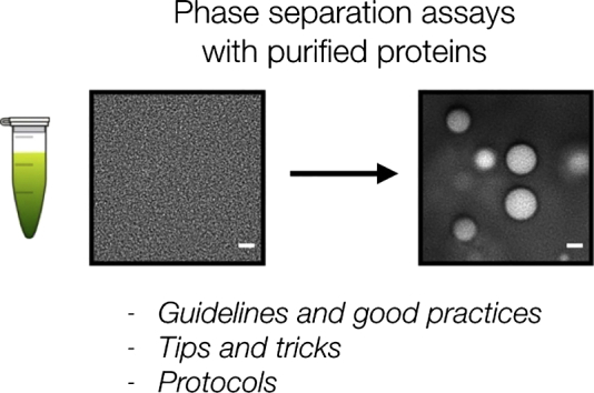
A User's Guide for Phase Separation Assays with Purified Proteins
- PMID: 29944854
- PMCID: PMC6215329
- DOI: 10.1016/j.jmb.2018.06.038
A User's Guide for Phase Separation Assays with Purified Proteins
Abstract
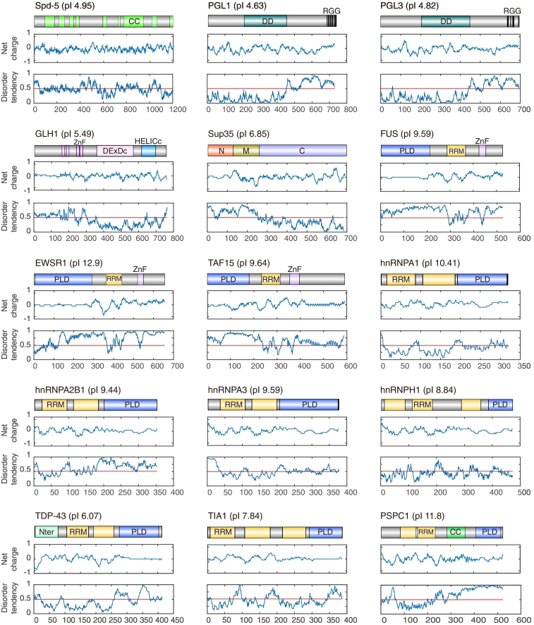
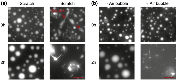
The formation of membrane-less organelles and compartments by protein phase separation is an important way in which cells organize their cytoplasm and nucleoplasm. In vitro phase separation assays with purified proteins have become the standard way to investigate proteins that form membrane-less compartments. By now, various proteins have been purified and tested for their ability to phase separate and form liquid condensates in vitro. However, phase-separating proteins are often aggregation-prone and difficult to purify and handle. As a consequence, the results from phase separation assays often differ between labs and are not easily reproduced. Thus, there is an urgent need for high-quality proteins, standardized procedures, and generally agreed-upon practices for protein purification and conducting phase separation assays. This paper provides protocols for protein purification and guides the user through the practicalities of in vitro protein phase separation assays, including best-practice approaches and pitfalls to avoid. We believe that this compendium of protocols and practices will provide a useful resource for scientists studying the phase behavior of proteins.
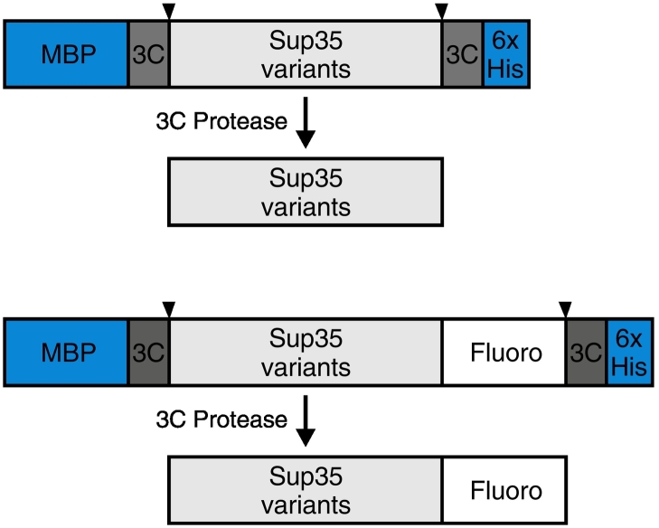
Keywords: Sup35; low-complexity proteins; membrane-less compartment; membrane-less organelle; phase separation; prion-like protein; protein purification.
Copyright © 2018 The Authors. Published by Elsevier Ltd.. All rights reserved.
Figures

References
Publication types
MeSH terms
Substances
LinkOut - more resources
Full Text Sources
Other Literature Sources
