. 2018 Jun 21;70(6):1067-1080.e12.
doi: 10.1016/j.molcel.2018.04.022. Epub 2018 Jun 6.

The Initial Response of a Eukaryotic Replisome to DNA Damage

Affiliations

The Initial Response of a Eukaryotic Replisome to DNA Damage

Martin R G Taylor et al. Mol Cell. .

Abstract

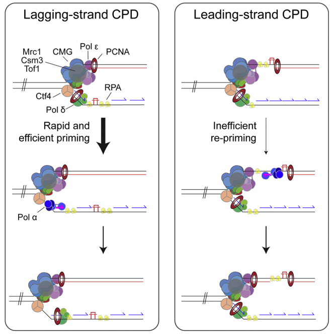
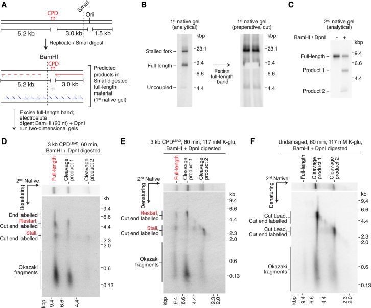
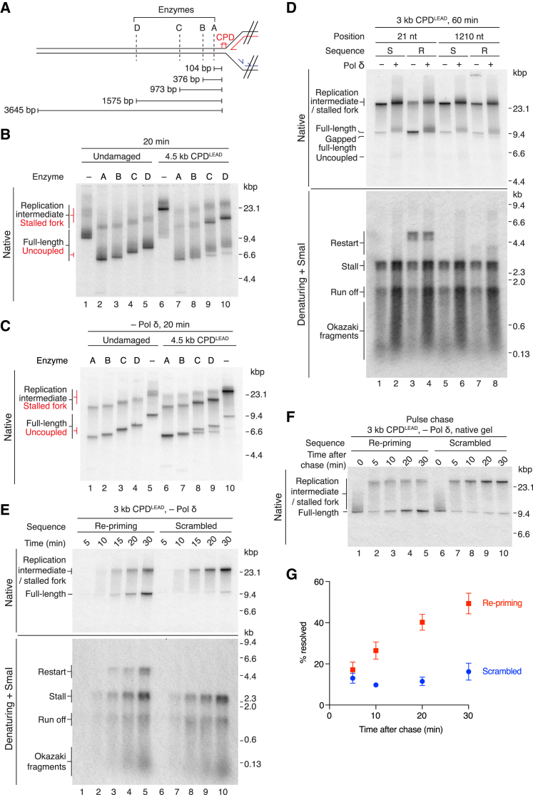
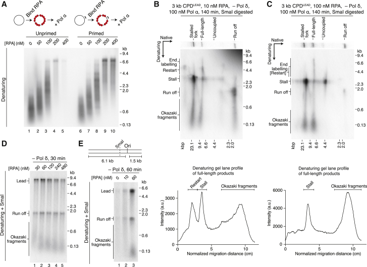
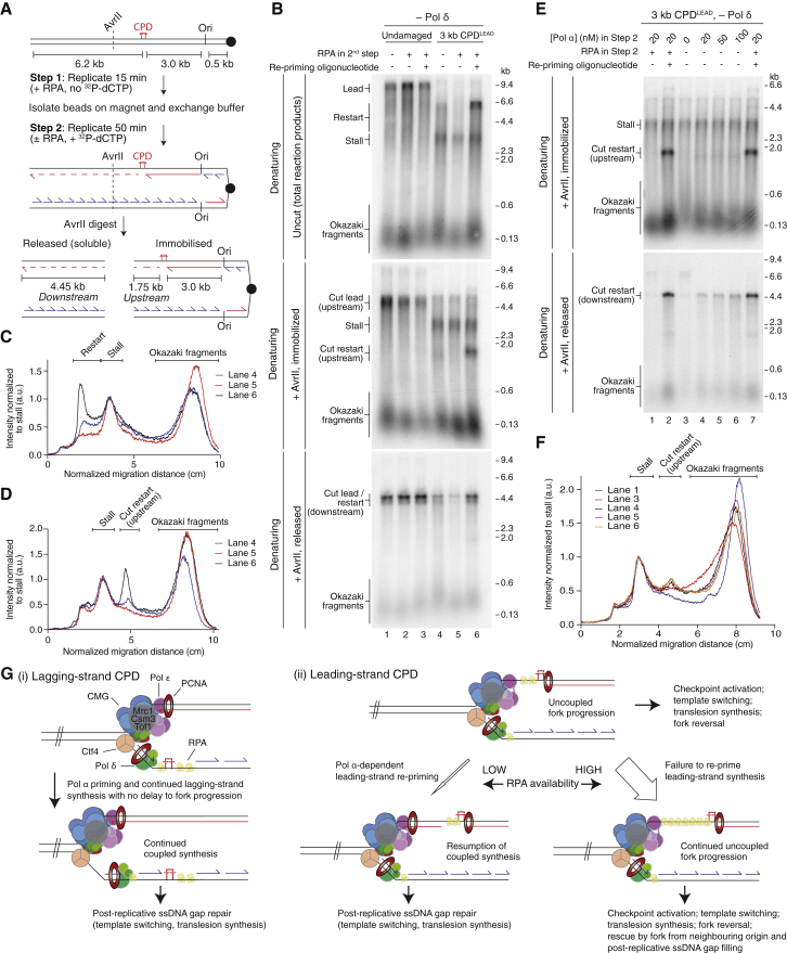
The replisome must overcome DNA damage to ensure complete chromosome replication. Here, we describe the earliest events in this process by reconstituting collisions between a eukaryotic replisome, assembled with purified proteins, and DNA damage. Lagging-strand lesions are bypassed without delay, leaving daughter-strand gaps roughly the size of an Okazaki fragment. In contrast, leading-strand polymerase stalling significantly impacts replication fork progression. We reveal that the core replisome itself can bypass leading-strand damage by re-priming synthesis beyond it. Surprisingly, this restart activity is rare, mainly due to inefficient leading-strand re-priming, rather than single-stranded DNA exposure or primer extension. We find several unanticipated mechanistic distinctions between leading- and lagging-strand priming that we propose control the replisome's initial response to DNA damage. Notably, leading-strand restart was specifically stimulated by RPA depletion, which can occur under conditions of replication stress. Our results have implications for pathway choice at stalled forks and priming at DNA replication origins.

Keywords: DNA; DNA damage response; DNA damage tolerance; DNA repair; DNA replication; genome stability; primase; re-priming; replication fork; replisome.


Figures

None
Graphical abstract
Figure 1
Figure 1
The Replisome Rapidly and Efficiently Bypasses a Lagging-Strand CPD (A) Schematic of AhdI- and BamHI-linearized undamaged templates and the predicted replication products. In this and all subsequent figures; red: leading-strands; blue: lagging-strands; the position of the ARS306 origin of replication is marked, Ori. The location of the SmaI restriction site is indicated. (B) Standard replication reactions performed on the templates illustrated in (A). Unless stated otherwise, this and all subsequent standard replication assays contained 217 mM potassium glutamate. Templates were prepared by linearizing maxiprep DNA (Maxi) or undamaged plasmids prepared using the same method used to generate CPD containing plasmids (Ligated). (C) Schematic of the 6.7 kb CPDLAG template and the predicted replication products of lagging-strand lesion bypass. (D) Replication reaction comparing undamaged and 6.7 kb CPDLAG templates. (E) Schematic of the 5.1 kb CPDLAG template. (F) Pulse-chase experiment on undamaged and 5.1 kb CPDLAG templates. The chase was added at 2 min 50 s. (G) Quantitation of pulse-chase experiments performed as in (F). Error bars represent the SEM from four experiments. Data were fit to a linear regression. Dashed line indicates the distance from Ori to CPDLAG (5.1 kb).
Figure 2
Figure 2
Daughter-Strand Gaps Are Generated during Bypass of CPDLAG (A) Schematic showing the location of restriction sites relative to the 5.1 kb CPDLAG. (B) Replication products from undamaged and 5.1 kb CPDLAG templates were digested post-replicatively. The distances from enzyme recognition sequences to the TT dinucleotide that is crosslinked in CPD templates are shown (cutting position). (C) Quantitation of full-length products from experiments performed as in (B). Error bars represent the SEM from three experiments. (D) Two-dimensional gel of post-replicatively digested CPDLAG products. Full-length products in the native gel are composed of run off products and Okazaki fragments. (E) Effect of Pol α concentration on daughter-strand gap size. Replication products were digested post-replicatively with AvrII. (F) Quantitation of full-length products from Pol α titrations performed as in (E). Error bars represent the SEM from three experiments.
Figure 3
Figure 3
Response of the Replisome to a Leading-Strand CPD (A) Schematic showing the 3 kb CPDLEAD template and the predicted replication products of leading-strand lesion bypass by re-priming. In this and all subsequent figures the putative restart product is shown as a dashed red line. (B and C) Comparison of replication products from undamaged and 3 kb CPDLEAD templates in the presence (B) and absence of Pol δ (C). (C) In this and all subsequent experiments entirely lacking Pol δ, the reaction buffer contained 117 mM potassium glutamate. (D) Two-dimensional gel of a replication reaction performed on the 3 kb CPDLEAD template (top), together with a schematic of the products generated (bottom). (E and F) Pulse-chase experiments on undamaged (E) and 4.5 kb CPDLEAD templates (F). The chase was added 14 min 50 s after replication was initiated. (G and H) Quantitation of pulse-chase experiments on undamaged (G) and 4.5 kb CPDLEAD templates (H) performed as in (E) and (F). “Resolved” is the sum of full-length and uncoupled products. Error bars represent the SEM from four experiments.
Figure 4
Figure 4
Evidence of Leading-Strand Re-priming by the Core Eukaryotic Replisome (A) Schematic of the 3 kb CPDLEAD template and the predicted replication products of leading-strand lesion bypass by re-priming followed by post-replicative SmaI digestion. (B) SmaI-digested replication products (60 min) were run in a native gel (analytical, 2%, one lane; preparative, 98%, two lanes). Full-length products were electroeluted from excised preparative lane gel slices. (C and D) Electroeluted DNA from (B) was digested with BamHI and DpnI and analyzed by native (C) and two-dimensional electrophoresis (BamHI + DpnI-digested sample) (D). (E and F) Electroeluted full-length products from CPDLEAD (E) and undamaged (F) templates in Figure S4C were digested with BamHI and DpnI and separated through two-dimensional gels. In (D) and (E), products arising from leading-strand re-priming are shown in red.
Figure 5
Figure 5
Priming Is the Main Limiting Step in Leading-Strand Restart (A) Schematic of the downstream region of the AhdI-linearized 4.5 kb CPDLEAD template and the positions of restriction sites used to truncate the template prior to replication. Distances are measured from the CPD to the nucleotide after which the first cut is made by the restriction enzyme. (B and C) Replication reactions performed on the templates illustrated in (A) in the presence (B) and absence (C) of Pol δ. Products specific to the CPDLEAD templates are annotated in red. (D) Replication of the 3 kb CPDLEAD template in the presence of re-priming (R) or scrambled (S) oligonucleotides. The distance from the CPD to the distal end of the re-priming oligonucleotide binding site is illustrated (position). (E and F) Time course (E) and pulse-chase experiments (F) in the presence of a re-priming (21 nt position) or scrambled oligonucleotide. (F) The chase was added at 4 min 50 s. (G) Quantitation of pulse-chase experiments as performed in (F). Error bars represent the SEM from three experiments.
Figure 6
Figure 6
RPA Levels Differentially Affect Lagging- and Leading-Strand Priming (A) Primase assay. RPA was pre-bound to unprimed (left) and primed (right) M13mp18 ssDNA for 10 min before addition of Pol α for 20 min. 120 nM RPA is saturating assuming a binding footprint of 30 nt. (B and C) Two-dimensional gels of replication assays performed on the 3 kb CPDLEAD template with 10 nM (B) or 100 nM (C) RPA. Lane profiles showing the constituents (denaturing) of the full-length products are shown below each gel. (D) RPA titration on an AhdI-linearized undamaged template. (E) RPA titration on a truncated undamaged template as illustrated.
Figure 7
Figure 7
RPA Depletion Stimulates Pol α-Dependent Leading-Strand Restart (A) Reaction scheme for bead-bound replication assays. (B–D) Reactions performed as illustrated in (A) on undamaged and 3 kb CPDLEAD templates (20 nM Pol α throughout). Products were separated through denaturing gels (B), and lane profiles for uncut (C) and immobilized AvrII-treated samples (D) are shown. (E and F) Reaction performed as illustrated in (A) but with 5 nM Pol α in step 1 and varying concentrations of Pol α in step 2 as indicated. AvrII digested products were separated though denaturing gels (E), and lane profiles for the immobilized samples (F) are shown. (G) Model of the initial response of the replisome to (i) lagging- and (ii) leading-strand CPDs.

References

    1. Bianchi J., Rudd S.G., Jozwiakowski S.K., Bailey L.J., Soura V., Taylor E., Stevanovic I., Green A.J., Stracker T.H., Lindsay H.D., Doherty A.J. PrimPol bypasses UV photoproducts during eukaryotic chromosomal DNA replication. Mol. Cell. 2013;52:566–573. - PMC - PubMed
    1. Biswas D., Yu Y., Prall M., Formosa T., Stillman D.J. The yeast FACT complex has a role in transcriptional initiation. Mol. Cell. Biol. 2005;25:5812–5822. - PMC - PubMed
    1. Collins K.L., Kelly T.J. Effects of T antigen and replication protein A on the initiation of DNA synthesis by DNA polymerase alpha-primase. Mol. Cell. Biol. 1991;11:2108–2115. - PMC - PubMed
    1. Coster G., Frigola J., Beuron F., Morris E.P., Diffley J.F. Origin licensing requires ATP binding and hydrolysis by the MCM replicative helicase. Mol. Cell. 2014;55:666–677. - PMC - PubMed
    1. Daigaku Y., Davies A.A., Ulrich H.D. Ubiquitin-dependent DNA damage bypass is separable from genome replication. Nature. 2010;465:951–955. - PMC - PubMed

Publication types

MeSH terms