. 2018 Sep;1862(9):1925-1932.
doi: 10.1016/j.bbagen.2018.06.015. Epub 2018 Jun 23.

Human cathepsins K, L, and S: Related proteases, but unique fibrinolytic activity

Affiliations

Human cathepsins K, L, and S: Related proteases, but unique fibrinolytic activity

Simone A Douglas et al. Biochim Biophys Acta Gen Subj. 2018 Sep.

Abstract

Background: Fibrin formation and dissolution are attributed to cascades of protease activation concluding with thrombin activation, and plasmin proteolysis for fibrin breakdown. Cysteine cathepsins are powerful proteases secreted by endothelial cells and others during cardiovascular disease and diabetes. Their fibrinolytic activity and putative role in hemostasis has not been well described.

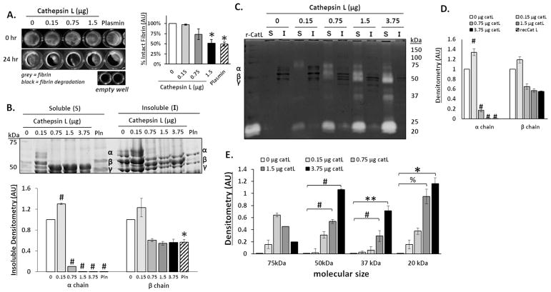
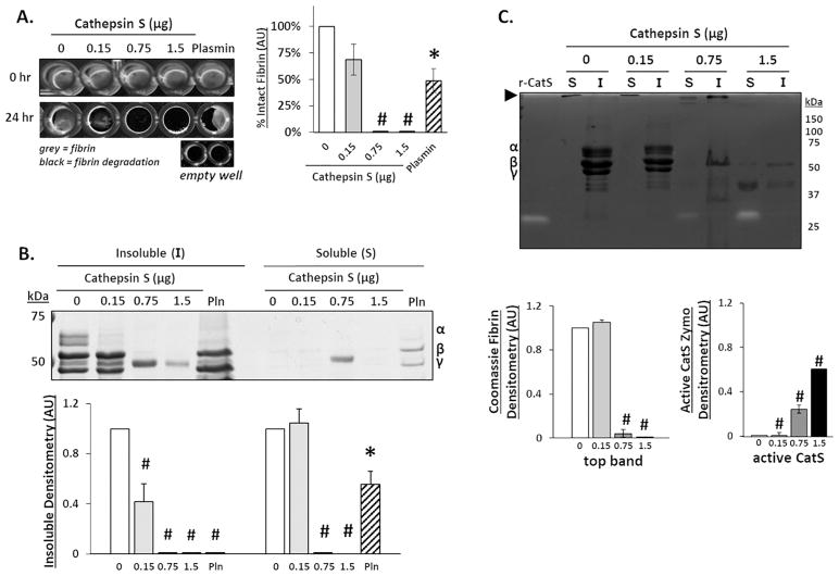
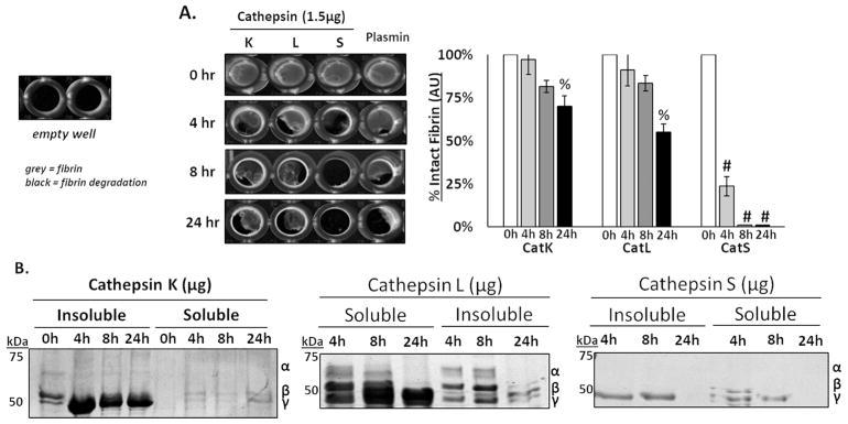
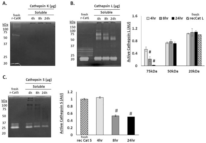
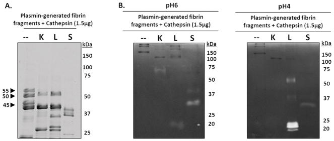
Methods: Fibrin gels were polymerized and incubated with recombinant human cathepsins (cat) K, L, or S, or plasmin, for dose-dependent and time-dependent studies. Dissolution of fibrin gels was imaged. SDS-PAGE was used to resolve cleaved fragments released from fibrin gels and remnant insoluble fibrin gel that was solubilized prior to electrophoresis to assess fibrin α, β, and γ polypeptide hydrolysis by cathepsins. Multiplex cathepsin zymography determined active amounts of cathepsins remaining.

Results: There was significant loss of α and β fibrin polypeptides after incubation with cathepsins, with catS completely dissolving fibrin gel by 24 h. Binding to fibrin stabilized catL active time; it associated with cleaved fibrin fragments of multiple sizes. This was not observed for catK or S. CatS also remained active for longer times during fibrin incubation, but its association/binding did not withstand SDS-PAGE preparation.

Conclusions: Human cathepsins K, L, and S are fibrinolytic, and specifically can degrade the α and β fibrin polypeptide chains, generating fragments unique from plasmin.

General significance: Demonstration of cathepsins K, L, and S fibrinolytic activity leads to further investigation of contributory roles in disrupting vascular hemostasis, or breakdown of fibrin-based engineered vascular constructs where non-plasmin mediated fibrinolysis must be considered.

Keywords: Biomaterials; Cathepsins; Clotting; Fibrinolysis; Hemostasis; Proteases.


Figures

Figure 1
Figure 1. Cathepsin L degrades fibrin gels, releasing fibrin degradation products into the supernatant
Fibrin gels (100 μl) were polymerized in 96 well plates then incubated with increasing amounts of cathepsin L (catL) in 50 μl assay buffer for 24hrs. (A) Fibrin gels were imaged in 96 well plates before (0 h) and after (24 h) incubation, where grey color indicates intact fibrin and black indicates areas of degraded fibrin gel, as shown by the black levels in the empty wells. Area of grey intensity were quantified to determine intact fibrin percentage. The fibrin gel was degraded by 3%, 26%, and 48% for 0.15, 0.75, and 1.5 μg, respectively, with a significant decrease at 1.5μg of catL compared to the no enzyme control. (B) After degradation, the fibrin fragments released into the supernatant (denoted to as the soluble fraction) were collected and the remaining fibrin gel (insoluble fraction), was collected and solubilized with β-mercaptoethanol to prepare it for SDS-PAGE. Coomassie stained protein bands of α (63.5 kDa), β (56 kDa), and γ (47 kDa) chains of fibrin are visible in the insoluble fraction. Densitometry of bands are quantified in the graph below. As the amount of catL increased, there was a significant loss of the α chain from the insoluble gel fraction. (C) Cathepsin zymography was used to assess the amount of active catL. Active catL bands were identified in the soluble fraction at multiple molecular sizes. Unbound active catL appears at 20 kDa and the higher molecular weight (75 to 37kDa) bands of active catL appear in a cascading pattern that corresponds to the degraded α and β fibrin fragments. D) Densitometry of insoluble α and β chains of fibrin bands visible in the zymogram (darker bands) are quantified in the graph. E) Active catL bands (cleared white bands) at 75 kDa, 50 kDa, 37 kDa, and 20 kDa bands are quantified by densitometry in the graph (n=3–5, **p<0.05; %p<0.01; *p<0.005; # p<0.0001).
Figure 2
Figure 2. Cathepsin K degrades α and β fibrin polypeptide chains
Increasing amounts of cathepsin K (catK) were incubated with fibrin gels for 24 hours in 96-well plates. (A) Fibrin gels were imaged before and after incubation, with grey indicating intact fibrin. As the amount of catK increased, more fibrin degradation was observed with a significant decrease in fibrin with 1.5μg of catK. (B) Loss of fibrin α, β, and γ bands in the insoluble gel was determined using SDS-PAGE; densitometry below is for the α and β chain of remaining fibrin gel (insoluble). In the soluble fraction, little to no fibrin fragments were detected. (C) The zymogram showed no active catK remained in either the fibrin gel or in the supernatant with the released fibrin fragments. (n=5, *p<0.005; ^ p<0.001, # p<0.0001)
Figure 3
Figure 3. Cathepsin S degrades the α, β, and γ fibrin polypeptide chains, and fibrin fragments
Fibrin gels were formed and incubated with increasing amounts of cathepsin S (catS) for 24 hours. (A) 96-well plates were imaged pre- and post cathepsin incubation. As the catS amount increased, more fibrin degradation was observed and was quantified as shown with complete degradation of fibrin with 0.75 and 1.5μg of catS. (B) SDS-PAGE used to image loss of fibrin α, β, and γ polypeptides due to catS hydrolysis fibrin degradation; densitometry below is for the α and β chain of remaining fibrin gel (insoluble) normalized to the no enzyme control. Increased catS amounts correlated with loss of the α and β fibrin chains in the fibrin gel (insoluble fraction) and little to no detection of fibrin fragments (soluble fraction). Densitometry is shown below for the α and β fibrin gel. (C) Zymography was used to quantify active catS. Unbound catS was detected in the soluble fraction with increasing intensity correlated to increasing catS amount. Large macromolecules that are unable to migrate through the gel are also observed in the soluble fraction (open arrow at top of gel image). (n=5, *p<0.05; #p<0.0001).
Figure 4
Figure 4. Time course of fibrin gel degradation by cathepsins K, L, and S
Fibrin gels were incubated with 1.5 μg of cathepsins K, L, and S (cat K, L and S), and reactions were stopped after 4, 8, and 24 hours. (A) 96-well plate imaged, where grey represents intact fibrin and black represents areas of fibrin degradation. As time increased, there was a significant loss of fibrin gel between 4 and 24 hours of degradation by catK, L, and S. (B) CatS degraded fibrin the fastest, where the α and β chains of fibrin degraded by as little as 4 hours. 0 hour control time points for insoluble and soluble fractions shown in catK gel. (n=5, % p<0.01; #p<0.0001).
Figure 5
Figure 5. Cathepsins L and S remain active over longer periods of time in the presence of fibrin
Zymography was used to determine the ability for fibrin to extend the activity time of cathepsins K, L, and S. (A) No active catK was detected in the supernatant after 4, 8, or 24 hours. (B) Bands of active catL appear with a cascading loss of higher molecular weight bands (100, 75, 50, and 37kDa) between 4 and 24 hours. (C) Active catS was only detected at 25kDa, its expected molecular weight, with the amount of active catS decreasing over 24 hours. (n=4, #p<0.0001)
Figure 6
Figure 6. Cathepsins K, L, and S degrade plasmin-generated fibrin fragments
To identify if cathepsins K, L, or S could successively degrade the released fibrin fragments into smaller molecular weight fibrin fragments, fibrin gels were first incubated with plasmin for 24 hours, then the fibrin fragments released into the supernatant were collected then incubated with catK, L, or S for 24 hours. (A) Compared to the no enzyme control, catK, L, and S successively degraded the fibrin fragments into lower molecular weight fragments. CatS completely degraded the 55kDa, 50kDa, and 45kDa fragments while catK and L only degraded the 55kDa fibrin fragments. (B) From zymography, active catL was at its expected molecular size (20kDa), as well as the higher molecular weights (~50kDa and faintly at 37kDa), corresponding to the results from the dosing and time course fibrin degradation experiments. Active catK is observed at 75kDa. Unbound active catS was observed at 25kDa in the zymograms incubated at pH 6. Active catL appears in the zymograms incubated at pH 4.

Similar articles

Cited by

References

    1. Herrick S, Blanc-Brude O, Gray A, Laurent G. Fibrinogen. The International Journal of Biochemistry & Cell Biology. 1999;31:741–746. - PubMed
    1. Weisel JW, Veklich Y, Gorkun O. The Sequence of Cleavage of Fibrinopeptides from Fibrinogen is Important for Protofibril Formation and Enhancement of Lateral Aggregation in Fibrin Clots. Journal of Molecular Biology. 1993;232:285–297. - PubMed
    1. Doolittle RF. Fibrinogen and fibrin. Annu Rev Biochem. 1984;53:195–229. - PubMed
    1. Standeven KF, Ariens RA, Grant PJ. The molecular physiology and pathology of fibrin structure/function. Blood Rev. 2005;19:275–288. - PubMed
    1. Sloane MMM, Bonnie F. Cysteine cathepsins: multifunctional enzymes in cancer. Nature Reviews Cancer. 2006;6:764–775. - PubMed

Publication types

LinkOut - more resources