. 2018 Aug 1;141(8):2457-2474.
doi: 10.1093/brain/awy159.

Trisomy of human chromosome 21 enhances amyloid-β deposition independently of an extra copy of APP

Collaborators, Affiliations

Trisomy of human chromosome 21 enhances amyloid-β deposition independently of an extra copy of APP

Frances K Wiseman et al. Brain. .

Erratum in

  • Corrigendum.
    [No authors listed] [No authors listed] Brain. 2019 Jun 1;142(6):e25. doi: 10.1093/brain/awz061. Brain. 2019. PMID: 30879033 Free PMC article. No abstract available.

Abstract

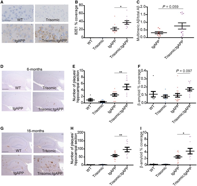
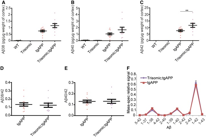
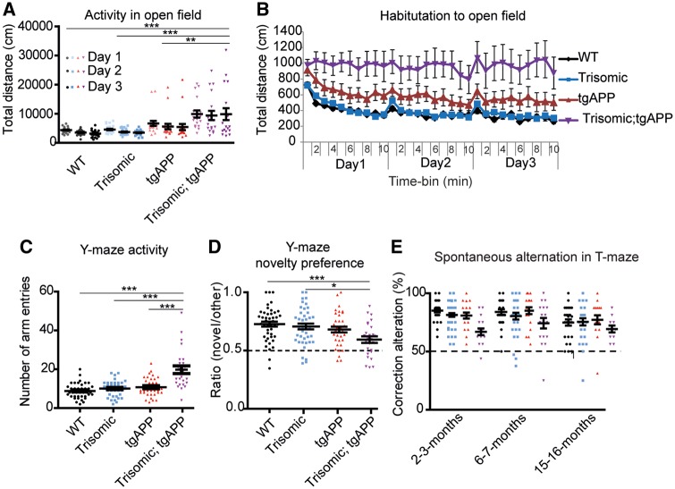
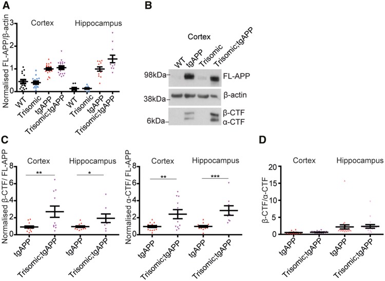
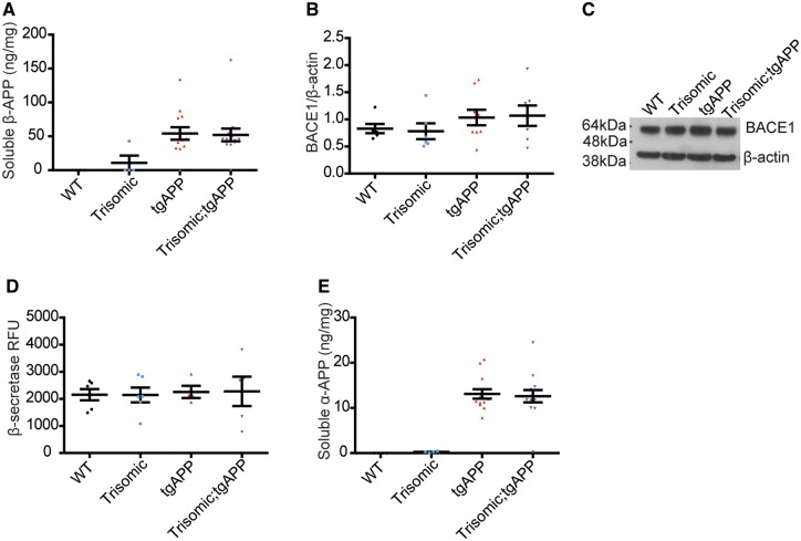
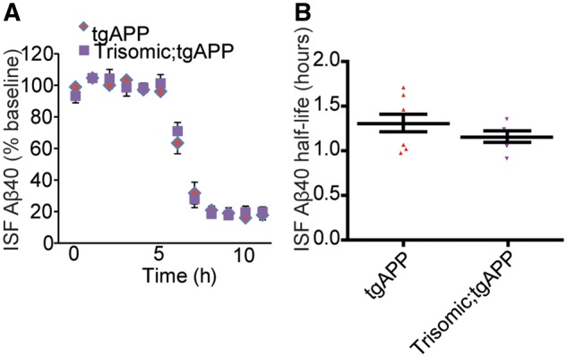
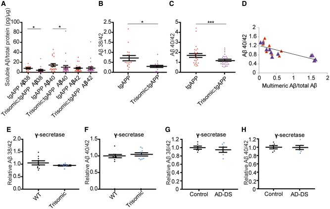
Down syndrome, caused by trisomy of chromosome 21, is the single most common risk factor for early-onset Alzheimer's disease. Worldwide approximately 6 million people have Down syndrome, and all these individuals will develop the hallmark amyloid plaques and neurofibrillary tangles of Alzheimer's disease by the age of 40 and the vast majority will go on to develop dementia. Triplication of APP, a gene on chromosome 21, is sufficient to cause early-onset Alzheimer's disease in the absence of Down syndrome. However, whether triplication of other chromosome 21 genes influences disease pathogenesis in the context of Down syndrome is unclear. Here we show, in a mouse model, that triplication of chromosome 21 genes other than APP increases amyloid-β aggregation, deposition of amyloid-β plaques and worsens associated cognitive deficits. This indicates that triplication of chromosome 21 genes other than APP is likely to have an important role to play in Alzheimer's disease pathogenesis in individuals who have Down syndrome. We go on to show that the effect of trisomy of chromosome 21 on amyloid-β aggregation correlates with an unexpected shift in soluble amyloid-β 40/42 ratio. This alteration in amyloid-β isoform ratio occurs independently of a change in the carboxypeptidase activity of the γ-secretase complex, which cleaves the peptide from APP, or the rate of extracellular clearance of amyloid-β. These new mechanistic insights into the role of triplication of genes on chromosome 21, other than APP, in the development of Alzheimer's disease in individuals who have Down syndrome may have implications for the treatment of this common cause of neurodegeneration.


Figures

Figure 1
Figure 1
Trisomy of chromosome 21 promotes the intracellular and extracellular deposition of amyloid-β. (A and B) Intracellular amyloid-β deposition (82E1) was increased by trisomy in hippocampal CA3 pyramidal neurons in 4.5–6.5-month-old mice [ANOVA trisomy-tgAPP interaction F(1,31) = 5.125, P = 0.031] [Bonferroni pairwise comparisons trisomic;tgAPP with tgAPP P = 0.012; wild-type (WT) (black circles) n = 10, trisomic (blue squares) n = 10, tgAPP (red triangles) n = 10, trisomic;tgAPP (purple inverted triangles) n = 10]. (C) A trend for increased hippocampal Tris-soluble multimeric amyloid-β (82E1-82E1 ELISA) normalized to the sum of amyloid-β38, amyloid-β40 and amyloid-β42 in trisomic;tgAPP was observed [trisomy F(1,24) = 3.928, P = 0.059; tgAPP n = 8, trisomic;tgAPP n = 11)]. (DI) Amyloid-β deposition (6F/3D) in the hippocampus was quantified at (DF) 6 and (GI) 16 months of age. Trisomy increases (E and H) the number of plaques [tgAPP–trisomy interaction F(1,77) = 6.744, P = 0.011, Bonferroni pairwise comparisons trisomic;tgAPP with tgAPP 6 months P = 0.008, 16 months P = 0.003] and (F and I) the area covered by amyloid-β [tgAPP–trisomy interaction F(1,85) = 4.005, P = 0.049, Bonferroni pairwise comparisons trisomic;tgAPP with tgAPP 6 months P = 0.097, 16 months P = 0.037] (6 months wild-type n = 8, trisomic = 8, tgAPP n = 15, trisomic;tgAPP n = 13; 16 months wild-type n = 16, trisomic = 9, tgAPP n = 13, trisomic;tgAPP n = 11). Data are represented as mean ± SEM, *P < 0.05, **P < 0.01, ***P < 0.001. Both male and female mice were studied and sex was included as a factor in the ANOVA. Scale bar in A = 50 µm; D and G = 500 µm. Aβ = amyloid-β.
Figure 2
Figure 2
Trisomy of chromosome 21 promotes the aggregation of amyloid-β. (AF) Cortical proteins from 16-month-old mice were fractionated and 5 M guanidine hydrochloride soluble amyloid-β quantified by (AE) Meso Scale Discovery Assay or (F) mass spectrometry. (A and B) No effect of trisomy on amyloid-β38 [tgAPP-trisomy interaction F(1,28) = 0.385, P = 0.540] or amyloid-β40 [tgAPP-trisomy interaction F(1,28) = 0.962, P = 0.355] was observed, (C) but significantly more amyloid-β42 was detected in trisomic;tgAPP mice [tgAPP-trisomy interaction F(1,28) = 5.573, P = 0.025] (Bonferroni pairwise comparisons trisomic;tgAPP with tgAPP P = 0.005, wild-type n = 6, trisomic = 6, tgAPP n = 13, trisomic;tgAPP n = 11). (D and E) No change in the amyloid-β38/42 ratio [trisomy F(1,19) = 0.072, P = 0.792] or amyloid-β40/42 ratio [trisomy F(1,19) = 0.047, P = 0.831] was observed. (F) The relative abundance of different forms of amyloid-β peptides was not altered by trisomy of chromosome 21 (tgAPP n = 12, trisomic;tgAPP n = 10). Data are represented as mean ± SEM, *P < 0.05, **P < 0.01, ***P < 0.001. Both male and female mice were used and sex was included as a factor in the ANOVA. Aβ = amyloid-β; WT = wild-type.
Figure 3
Figure 3
Trisomy of chromosome 21 exacerbates APP/amyloid-β-associated cognitive deficits. (A and B) Exposure to a novel open field was used as a test of activity and habituation (6 months, wild-type n = 28, trisomic n = 28, TgAPP n = 20, trisomic;tgAPP n = 18). (A) Overall activity: ANOVA of distance travelled revealed a main effect of tgAPP [F(1,78) = 26.250, P < 0.001], trisomy [F(1,78) = 9.246, P = 0.003], and a tgAPP-trisomy interaction [F(1,78) = 7.818, P = 0.007] (Bonferroni pairwise comparisons trisomic;tgAPP with wild-type P < 0.001, trisomic P < 0.001, and tgAPP P = 0.008). (B) The total distance moved declined with exposure time (1-min time bins) ANOVA: main effect of time bin [F(29,2262) = 12.399 P < 0.001]; an interaction of time bin × trisomy [ANOVA F(29,2262) = 1.789 P = 0.006], time bin × tgAPP [F(29,2262) = 1.560 P = 0.029] and time bin × trisomy;tgAPP [F(29,2262) = 1.983, P < 0.001] was observed by ANOVA. (C and D) A Y-maze spatial novelty preference task (1-min delay) was used as a test of activity and memory. (Cohort B 2–3 months and Cohort A 6–7 months, the effect of genotype was similar in both cohorts so data were combined for analysis, wild-type n = 45, trisomic n = 43, TgAPP n = 36, trisomic;tgAPP n = 26). (C) ANOVA of the number of arm entries (test phase), revealed a main effect of trisomy [F(1,89) = 50.360, P < 0.001], tgAPP [F(1,89) = 47.001, P < 0.001], and a tgAPP–trisomy interaction [F(1,89) = 31.720, P < 0.001] (Bonferroni pairwise comparisons trisomic;tgAPP with wild-type P < 0.001, trisomic P < 0.001, and tgAPP P < 0.001). (D) A preference ratio of 0.5 indicates chance performance (black dotted line). ANOVA of novelty preference revealed a main effect of trisomy [F(1,89) = 10.144 P = 0.002], tgAPP [F(1,89) = 9.312 P = 0.003] and a tgAPP–trisomy interaction [F(1,89) = 5.736, P = 0.019] (Bonferroni pairwise comparisons trisomic;tgAPP with wild-type P < 0.001 and trisomic P = 0.010). Performance of tgAPP–trisomic mice was above chance (one-sample t-test t = 3.287 P < 0.001). (E) A discrete-trial, longitudinal spontaneous alternation task in a T-maze was used as a test of memory, 50% alternation represents chance performance (black dotted line) (2–3 months wild-type n = 29, trisomic n = 30, TgAPP n = 21, trisomic;tgAPP n = 17, 6–7 months wild-type n = 28, trisomic n = 29, TgAPP n = 20, trisomic;tgAPP n = 17, 15–16 months wld-type n = 27, trisomic n = 26, TgAPP n = 27, trisomic;tgAPP n = 11). ANOVA of alternation showed a main effect of trisomy [F(1,67) = 7.084 P = 0.010], and an interaction of tgAPP–trisomy [F(1,67) = 4.706, P = 0.034] (Bonferroni pairwise comparison trisomic;tgAPP with wild-type P = 0.032). Performance of tgAPP-trisomic mice was above chance (one-sample t-test, 2–3 months t = 5.884 P < 0.001, 6–7 months t = 5.378 P < 0.001, 15–16 months t = 6.495 P < 0.001). Data are represented as mean ± SEM, *P < 0.05, **P < 0.01, ***P < 0.001. Both male and female mice were used and sex was included as a variable in the ANOVA. Aβ = amyloid-β; WT = wild-type.
Figure 4
Figure 4
Trisomy of chromosome 21 genes other than APP does not increase APP abundance nor alter β-CTF/α-CTF ratio. (A, B and D) Full-length APP (FL-APP), APP β-CTF and APP α-CTF were measured in cortex (wild-type n = 17, trisomic n = 16, tgAPP n = 24, trisomic;tgAPP n = 19) and hippocampus (wild-type n = 11, trisomic n = 12, tgAPP n = 24, trisomic;tgAPP n = 17) at 3 months of age. (A) Full-length APP was higher in tgAPP and trisomic;tgAPP compared with wild-type or trisomic mice [cortex F(1,68) = 87.667, P < 0.001, hippocampus F(1,56) = 94.301, P < 0.001]. Trisomy did not alter full-length APP [trisomy–tgAPP interaction, cortex F(1,68) = 0.483, P = 0.489, hippocampus F(1,56) = 2.457, P = 0.123]. (B and C) In male mice, APP-CTF/full-length APP ratio was altered (cortex tgAPP n = 17, trisomic;tgAPP n = 11, hippocampus tgAPP n = 14, trisomic;tgAPP n = 8) β-CTF/full-length APP (t-test cortex P = 0.005, hippocampus P = 0.0217) and α-CTF/full-length APP (t-test cortex P = 0.005 hippocampus P < 0.001). (D) Trisomy did not alter the β-CTF/α-CTF ratio in the cortex [trisomy F(1,37) = 0.065, P = 0.799] or hippocampus [trisomy F(1,37) = 1.082, P = 0.305]. (B) Cropped western blot, four lanes of an eight-lane gel. Data are represented as mean ± SEM, *P < 0.05, **P < 0.01, ***P < 0.001. WT = wild-type.
Figure 5
Figure 5
Trisomy of chromosome 21 genes other than APP does not alter α-/β-secretase activity. Trisomy did not alter (A) soluble β-APP abundance [trisomy F(1,24) = 0.790, P = 0.383, trisomy-tgAPP interaction F(1,24) = 0.773, P = 0.388, wild-type n = 4, trisomic n = 4, TgAPP n = 13, trisomic;tgAPP n = 14], (B and C) cortical BACE1 abundance [trisomy F(1,24) = 0.002, P = 0.963; trisomy–tgAPP interaction F(1,24) = 0.071, P = 0.792, wild-type n = 6, trisomic n = 6, TgAPP n = 9, trisomic;tgAPP n = 7], (D) BACE1 β-secretase activity [trisomy F(1,13) = 0.006, P = 0.941; trisomy–tgAPP interaction F(1,13) = 0.001 P = 0.971, wild-type n = 6, trisomic n = 6, TgAPP n = 4, trisomic;tgAPP n = 5], or (E) soluble α-APP abundance [trisomy F(1,27) = 0.041, P = 0.841; trisomy–tgAPP interaction F(1,27) = 0.002, P = 0.969, wild-type n = 4, trisomic n = 4, TgAPP n = 13, trisomic;tgAPP n = 14]. (C) Cropped western blot, four lanes of an eight-lane gel. Data are represented as mean ± SEM. Both sexes were analysed and sex was included as a factor in the ANOVA. Aβ = amyloid-β.
Figure 6
Figure 6
Trisomy of chromosome 21 does not alter the half-life of extracellular amyloid-β. (A and B) The in vivo half-life of amyloid-β40, measured by microdialysis of hippocampal interstitial fluid (ISF), was not altered by trisomy of chromosome 21. Compound E injected at 6 h to halt further amyloid-β generation (t-test P = 0.258, tgAPP n = 8, trisomic;tgAPP n = 6 females only). Data are represented as mean ± SEM. Aβ = amyloid-β.
Figure 7
Figure 7
Trisomy of chromosome 21 modulates amyloid-β ratios in vivo independently of modulation of γ-secretase activity. (AC) Tris-soluble amyloid-β38, amyloid-β40 and amyloid-β42 were measured by Meso Scale Discovery 6E10 Aβ Triplex assay in 2-month-old hippocampus (wild-type n = 24, trisomic n = 22, TgAPP n = 25 and trisomic;tgAPP n = 27). (A) Trisomy decreased amyloid-β38 [trisomy F(1,4) = 15.403, P = 0.017] and amyloid-β40 [trisomy F(1,11) = 6.359, P = 0.028], but amyloid-β42 was not changed [trisomy F(1,11) = 2.978, P = 0.112], (B) resulting in an alteration in the amyloid-β38/42 ratio [trisomy F(1,4) = 14.553, P = 0.019] and (C) the amyloid-β40/42 ratio [trisomy F(1,10) = 95.694, P < 0.001]. (D) Hippocampal amyloid-β40/42 ratio negatively correlates with the relative abundance of aggregated amyloid-β (multimeric 82E1-82E1 amyloid-β ELISA) (linear correlation, R2 = 0.5485, P = 0.0003, tgAPP n = 8, trisomic;tgAPP n = 11). Trisomy did not alter the carboxypeptidase activity of the γ-secretase complex, as measured by (E and G) amyloid-β38/42 and (F and H) amyloid-β40/42 ratios produced in vitro by the complex isolated from cortex from (E and F) the Tc1 trisomic mouse [trisomy amyloid-β38/42 F(1,11) = 4.88, P = 0.499; amyloid-β42/40 F(1,11) = 0.799, P = 0.395, wild-type = 9, trisomic = 9] and (G and H) people with Down syndrome and Alzheimer’s disease (AD-DS) (n = 6) compared with age- and sex-matched individuals who did not have Down syndrome or dementia (control n = 6) [trisomy amyloid-β38/42 F(1,5) = 0.102, P = 0.763; amyloid-β42/40 F(1,5) = 0.187, P = 0.684]. (AF) Data are represented as mean ± SEM, (G and H) individual cases plotted, horizontal line indicates mean ± SEM, *P < 0.05, **P < 0.01, ***P < 0.001. Both male and females were studied and sex was included as a variable in the ANOVA. Aβ = amyloid-β; WT = wild-type.

Comment in

Similar articles

Cited by

References

    1. Ahmed RR, Holler CJ, Webb RL, Li F, Beckett TL, Murphy MP. BACE1 and BACE2 enzymatic activities in Alzheimer’s disease. J Neurochem 2010; 112: 1045–53. - PMC - PubMed
    1. Branca C, Shaw DM, Belfiore R, Gokhale V, Shaw AY, Foley C et al. . Dyrk1 inhibition improves Alzheimer’s disease-like pathology. Aging Cell 2017; 16: 1146–54. - PMC - PubMed
    1. Brault V, Pereira P, Duchon A, Herault Y. Modeling chromosomes in mouse to explore the function of genes, genomic disorders, and chromosomal organization. PLoS Genet 2006; 2: e86. - PMC - PubMed
    1. Castellano JM, Kim J, Stewart FR, Jiang H, DeMattos RB, Patterson BW et al. . Human apoE isoforms differentially regulate brain amyloid-beta peptide clearance. Sci Transl Med 2011; 3: 89ra57. - PMC - PubMed
    1. Cataldo AM, Petanceska S, Terio NB, Peterhoff CM, Durham R, Mercken M et al. . Abeta localization in abnormal endosomes: association with earliest Abeta elevations in AD and Down syndrome. Neurobiol Aging 2004; 25: 1263–72. - PubMed

Publication types

Substances