. 2018 Aug 21;46(14):7250-7260.
doi: 10.1093/nar/gky582.

Convergent evolution of integration site selection upstream of tRNA genes by yeast and amoeba retrotransposons

Affiliations

Convergent evolution of integration site selection upstream of tRNA genes by yeast and amoeba retrotransposons

Eva Kling et al. Nucleic Acids Res. .

Abstract

Transposable elements amplify in genomes as selfish DNA elements and challenge host fitness because their intrinsic integration steps during mobilization can compromise genome integrity. In gene-dense genomes, transposable elements are notably under selection to avoid insertional mutagenesis of host protein-coding genes. We describe an example of convergent evolution in the distantly related amoebozoan Dictyostelium discoideum and the yeast Saccharomyces cerevisiae, in which the D. discoideum retrotransposon DGLT-A and the yeast Ty3 element developed different mechanisms to facilitate position-specific integration at similar sites upstream of tRNA genes. Transcription of tRNA genes by RNA polymerase III requires the transcription factor complexes TFIIIB and TFIIIC. Whereas Ty3 recognizes tRNA genes mainly through interactions of its integrase with TFIIIB subunits, the DGLT-A-encoded ribonuclease H contacts TFIIIC subunit Tfc4 at an interface that covers tetratricopeptide repeats (TPRs) 7 and 8. A major function of this interface is to connect TFIIIC subcomplexes τA and τB and to facilitate TFIIIB assembly. During the initiation of tRNA gene transcription τB is displaced from τA, which transiently exposes the TPR 7/8 surface of Tfc4 on τA. We propose that the DGLT-A intasome uses this binding site to obtain access to genomic DNA for integration during tRNA gene transcription.


Figures

Figure 1.
Figure 1.
Topology of the RNA polymerase III transcription complex and the LTR retrotransposons DGLT-A and Ty3. (A) Model of the TFIIIC/TFIIIB complex based on experiments in S. cerevisiae (26,27). TFIIIC consists of the subunits Tfc1 (τ95), Tfc3 (τ138), Tfc4 (τ131), Tfc6 (τ91), Tfc7 (τ55), and Tfc8 (τ60). The factor is assembled in two subcomplexes, τA and τB, which are mainly connected by the interaction of Tfc3 with Tfc4. Brf1, a subunit of TFIIIB, is recruited by interacting with Tfc4 and subsequently incorporates TBP into the complex. Bdp1 interacts with the complex only transiently during transcription initiation by binding to the same interface on Tfc4 that is bound by Tfc3. Note that Tfc4 and Tfc1 of the τA subcomplex are the only subunits of TFIIIC currently identified in the D. discoideum genome based on sequence homology. The intragenic promoter elements (A box and B box) of a tRNA gene are indicated. The preferred integration sites of DGLT-A and Ty3 are located ∼15 bp upstream of the first nucleotide of a mature tRNA. (B) Schematic presentations of DGLT-A and Ty3. In DGLT-A, one single ORF (GAG POL) encodes the entire protein machinery, whereas in Ty3 the GAG3 and POL3 genes are organized in two overlapping reading frames and translated as Gag3 and Gag3-Pol3 fusion protein by a +1 frameshift. LTR: long terminal repeat; GAG: group-specific antigen; PR: protease; RT: reverse transcriptase; RNH: ribonuclease H; IN: integrase; PB, tRNA primer binding site; PPu, polypurine tract.
Figure 2.
Figure 2.
Interaction of DGLT-A proteins and Tfc4. (A) Amino acid positions of the DGLT-A GAG-POL polyprotein are indicated as inserted into yeast two-hybrid vectors. The DGLT-A integrase (IN) is characterized by a catalytic core domain (CCD; DGLT-A1148–1313) containing a DDE catalytic triad, an N-terminal domain (NTD; DGLT-A994–1147) containing a typical HHCC zinc finger-like motif, and a C-terminal extension containing a conserved GPY/F motif. For yeast two-hybrid experiments the IN-NTD was further split into the N-terminal extension domain (NED; DGLT-A994–1067) and the zinc finger (ZF) region (DGLT-A1068–1147). See Supplementary Figure S1B for an alignment of DGLT-A and Ty3 IN sequences. (B) Schematic presentation of D. discoideum Tfc4. Positions of the TPRs are indicated by red boxes with numbers. The amino acid positions Tfc4 fragments used in yeast two-hybrid screenings are listed. (C) Results of yeast two-hybrid experiments testing BD-fused TPR 7–10 of Tfc4 (Tfc4446–608) against AD-fused DGLT-A proteins as indicated. (D) Pull-down experiment testing the binding of FLAG-tagged Tfc4 TPR 7–10 (FLAG-Tfc4446–608) to GFP-tagged DGLT-A RNH (GFP-RNH). Both proteins were expressed in bacteria and used as purified proteins. Input refers to the purified proteins and is used as markers. Pull-down left panel: FLAG- Tfc4446–608 was bound to anti-FLAG antibodies immobilized on magnetic beads. The loaded beads were incubated with GFP- RNH (DGLT-A733–879). Pull-down middle panel: FLAG-Tfc4446–608 was immobilized on anti-FLAG beads and incubated with GFP (first negative control). Pull-down right panel: Beads carrying anti-FLAG antibodies were incubated with GFP-RNH without prior loading of the FLAG-tagged bait protein (second negative control). After western blotting, the proteins were first stained with anti-GFP antibodies and then with anti-FLAG antibodies. The asterisks indicate prominent degradation products of GFP-RNH. (E) Similar pull-down experiment using FLAG- Tfc4446–608 as bait and GFP-tagged IN-NED (DGLT-A994–1067) as the binding partner.
Figure 3.
Figure 3.
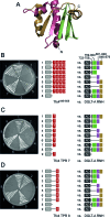
Interaction of DGLT-A RNH and IN-NTD with TFIIIC subunit Tfc4. (A) Solved structure of the central TPR arrays (TPRs 1–10) of yeast τTfc4. The picture was generated with PyMol based on PDB entry 5AIO (26). (B) Model of the central TPR region of D. discoideum Tfc4. The model was generated using the solved structure of the yeast Tfc4 TPRs 1–10 (PDB 5AIO) (26) as template. (C, D) Yeast two-hybrid assay testing different deletions of the right TPR arm (TPRs 7–10) against DGLT-A RNH (C) and DGLT-A IN-NTD (D).
Figure 4.
Figure 4.
Mapping of the DGLT-A RNH surface that binds to Tfc4 TPRs 7–10. (A) The model of the DGLT-A RNH domain was generated using the solved structure of RT-RNH of Ty3 (PDB entry 4OL8) (40) as template. The color code represents RNH fragments cloned for separate testing in the yeast two-hybrid system. (B) Yeast two-hybrid experiment testing for the interaction of the right TPR arrays of D. discoideum Tfc4 (TPRs7–10) with different parts of DGLT-A RNH. Note that positions 733–758 are localized outside of the RNH core domain and belong to the RT thumb region. (C, D) Similar yeast two-hybrid experiments using only TPR 7 and TPR 8 as baits.
Figure 5.
Figure 5.
Protein interactions within the DGLT-A element. (A) Yeast two-hybrid experiment localizing the surface on DGLT-A RNH required for binding to DGLT-A IN-NED. A comparison of IN-NTD with the corresponding part of yeast Ty3 IN-NTD is provided in Supplementary Figure S1. (B) Pull-down experiment to confirm the binding of DGLT-A IN-NED to DGLT-A RNH. Proteins were expressed in bacteria and used as purified proteins. Input refers to the purified proteins and is used as markers. Pull-down left panel: FLAG-tagged DGLT-A RNH (FLAG-DGLT-A733–879) was immobilized on anti-FLAG antibodies bound to magnetic beads. The loaded beads were incubated with GFP-tagged DGLT-A IN-NED (GFP-IN-NED containing DGLT-A994–1067). Pull-down middle panel: FLAG- RNH (DGLT-A733–879) was bound to anti-FLAG beads and incubated with GFP (first negative control). Pull-down right panel: Beads carrying anti-FLAG antibodies were incubated with GFP-IN-NED (DGLT-A994–1067) without prior loading of the FLAG-tagged bait protein (second negative control). The blotted proteins were first stained with anti-GFP antibodies and then with anti-FLAG antibodies. (C) Yeast two-hybrid experiment investigating dimerization of DGLT-A RNH. (D) Pull-down experiment testing GFP-tagged RNH against FLAG-tagged RNH (DGLT-A733–879). The asterisks indicate the prominent degradation products of GFP-RNH.
Figure 6.
Figure 6.
Comparison of interaction platforms on DGLT-A RNH. (A) The model of the RNH domain of DGLT-A is based on the solved structure of RT-RNH of Ty3 (PDB entry 4OL8). Mutations E771A, K831A, E864A at the surface of the β-sheets are presented as sticks. (B) The wild-type and triple mutant of DGLT-A RNH were tested for binding to Tfc4 (TPRs 7–10; Tfc4440–586), DGLT-A IN-NED (DGLT-A994–1067) and DGLT-A RNH (DGLT-A759–879). White asterisks indicate the triple mutant of RNH.
Figure 7.
Figure 7.
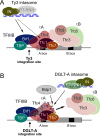
Model of integration site selection by Ty3 and DGLT-A. The τB complex contains the B box binding activity (Tfc3) and mediates the binding of TFIIIC (τA/τB complex) to tRNA genes. The assembly of TFIIIB is mediated by DNA-bound τA by interactions between TFC4 and TFIIIB subunit Brf1. Subsequently, TBP is incorporated primarily by interaction with Brf1 (25). (A) Model of Ty3 integration site selection. The Ty3 intasome (preintegration complex) consists at least of Ty3 IN and Ty3 cDNA. Note that it is not known whether RT/RNH is part of the Ty3 intasome. Ty3 selects tRNA genes by direct interaction of Ty3 IN with the TBP/Brf1 heterodimer (35,36). Ty3 IN also interacts with subunit Tfc1, which is not required for integration site selection but affects the orientation of integrated Ty3 copies relative to the target (37). (B) Model of target site selection by DGLT-A. After assembly of the TFIIIB Brf1–TBP complex, the τB complex is displaced by Bdp1 binding to the TPR8 region of Tfc4 (26). Both DGLT-A RNH and IN-NED target the TPR7/8 region of Tfc4, suggesting that the intasome of DGLT-A may compete with Bdp1 for binding on τA during the initiation of Pol III transcription. How the interaction of the DGLT-A intasome with the right TPR array of Tfc4 provides the integrase access to a DNA region immediately upstream of the transcription start site remains unknown.

Similar articles

Cited by

References

    1. Orgel L.E., Crick F.H.C.. Selfish DNA: the ultimate parasite. Nature. 1980; 284:604–607. - PubMed
    1. Doolittle W.F., Sapienza C.. Selfish genes, the phenotype paradigm and genome evolution. Nature. 1980; 284:601–603. - PubMed
    1. Sultana T., Zamborlini A., Cristofari G., Lesage P.. Integration site selection by retroviruses and transposable elements in eukaryotes. Nat. Rev. Genet. 2017; 18:292–308. - PubMed
    1. Kim J.M., Vanguri S., Boeke J.D., Gabriel A., Voytas D.F.. Transposable elements and genome organization: a comprehensive survey of retrotransposons revealed by the complete Saccharomyces cerevisiae genome sequence. Genome Res. 1998; 8:464–478. - PubMed
    1. Glöckner G., Szafranski K., Winckler T., Dingermann T., Quail M., Cox E., Eichinger L., Noegel A.A., Rosenthal A.. The complex repeats of Dictyostelium discoideum. Genome Res. 2001; 11:585–594. - PMC - PubMed

Publication types

MeSH terms

LinkOut - more resources