. 2018 Jul 17;145(14):dev164889.
doi: 10.1242/dev.164889.

SOX2 regulation by hedgehog signaling controls adult lingual epithelium homeostasis

Affiliations

SOX2 regulation by hedgehog signaling controls adult lingual epithelium homeostasis

David Castillo-Azofeifa et al. Development. .

Abstract

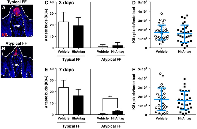
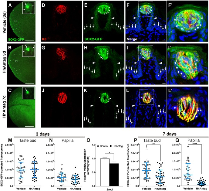
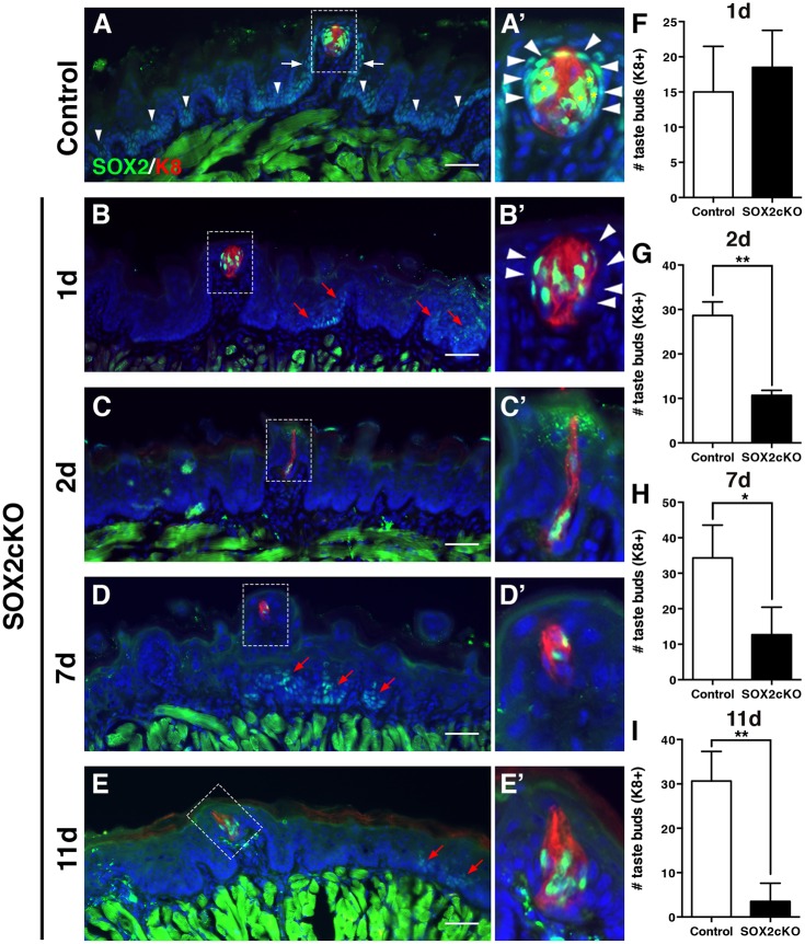
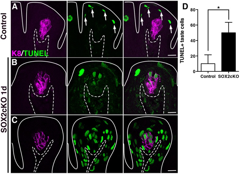
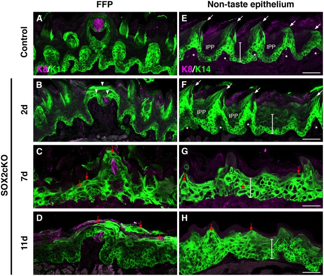
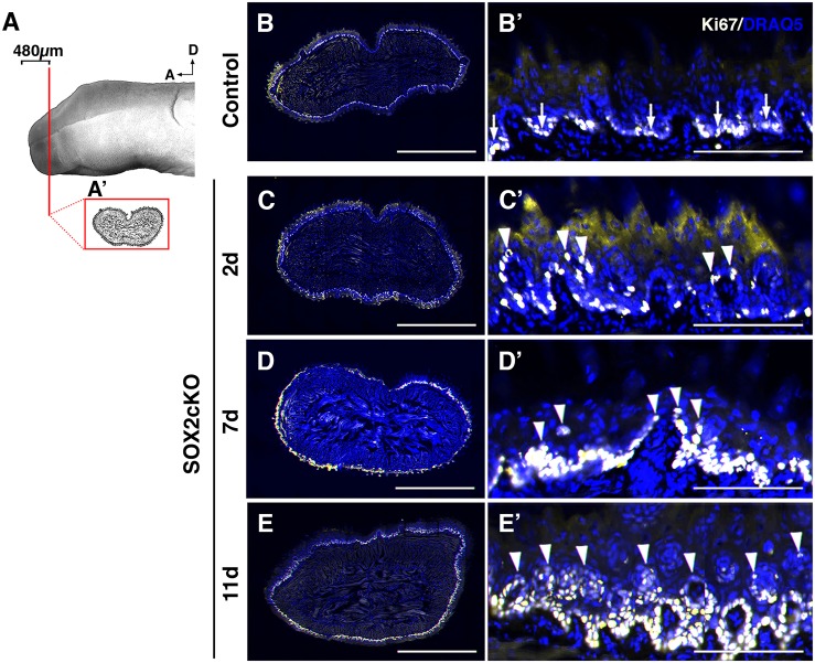
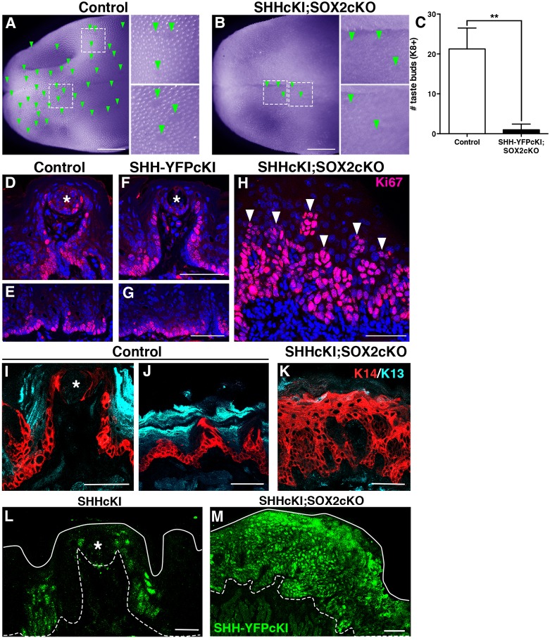
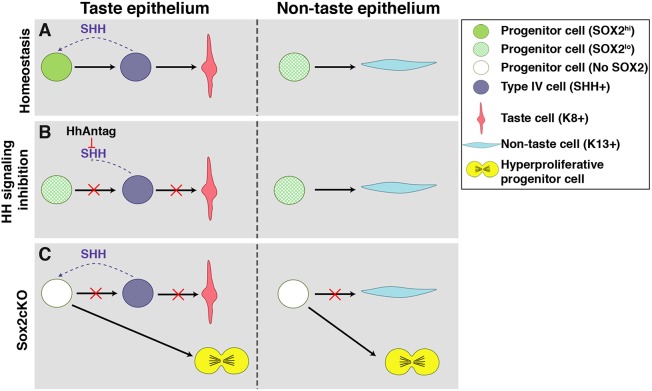
Adult tongue epithelium is continuously renewed from epithelial progenitor cells, a process that requires hedgehog (HH) signaling. In mice, pharmacological inhibition of the HH pathway causes taste bud loss within a few weeks. Previously, we demonstrated that sonic hedgehog (SHH) overexpression in lingual progenitors induces ectopic taste buds with locally increased SOX2 expression, suggesting that taste bud differentiation depends on SOX2 downstream of HH. To test this, we inhibited HH signaling in mice and observed a rapid decline in Sox2 and SOX2-GFP expression in taste epithelium. Upon conditional deletion of Sox2, differentiation of both taste and non-taste epithelial cells was blocked, and progenitor cell number increased. In contrast to basally restricted proliferation in controls, dividing cells were overabundant and spread to suprabasal epithelial layers in mutants. SOX2 loss in progenitors also led non-cell-autonomously to taste cell apoptosis, dramatically shortening taste cell lifespans. Finally, in tongues with conditional Sox2 deletion and SHH overexpression, ectopic and endogenous taste buds were not detectable; instead, progenitor hyperproliferation expanded throughout the lingual epithelium. In summary, we show that SOX2 functions downstream of HH signaling to regulate lingual epithelium homeostasis.

Keywords: Cell differentiation; Mouse; Progenitor proliferation; Smoothened antagonist; Taste bud.


Conflict of interest statement

Competing interestsThe authors declare no competing or financial interests.

Figures

Fig. 1.
Fig. 1.
Adult taste buds are mildly but significantly affected by 1 week of HhAntag treatment. (A,B) Although the morphology of typical FF (A) and atypical FF (B) papillae differ, both house K8+ taste buds (red). Images are confocal compressed z-stacks. Nuclei are counterstained with Draq5 (blue); dashed lines delimit the basement membrane; solid lines delimit the epithelial surface; mc, mesenchymal core. Scale bars: 10 μm.  (C) After 3 days of HhAntag treatment, typical FF and atypical FF taste bud numbers do not differ from vehicle-treated controls. (D) The number of K8+ pixels, i.e. taste bud size, is comparable between vehicle and HhAntag-treated mice at 3 days. (E) After 7 days, HhAntag-treated mice tend to have fewer typical FF taste buds, whereas atypical FF taste buds are significantly increased. (F) At 7 days, taste bud size does not differ between HhAntag-treated and control mice. N=3 mice for 3 days; N=3-4 mice for 7 days; n=30 taste buds for vehicle or 30-40 for HhAntag (ten taste buds randomly selected per mouse). Data are represented as mean±s.d.; **P<0.01 (Student's t-test).
Fig. 2.
Fig. 2.
SOX2-GFP expression is reduced in taste buds and papillae in mice treated with HhAntag. (A) In whole-mount preparations of vehicle-treated tongues at 3 days, taste buds appear to have high SOX2-GFP expression (inset, arrow), whereas adjacent FF papilla walls express lower SOX2-GFP (inset, arrowheads). (B) After 3 days of HhAntag treatment, a similar pattern of bright GFP expression in taste buds (inset, arrow) with dimmer GFP expression in papilla epithelium (inset, arrowheads) is evident. (C) After 7 days of HhAntag treatment, fewer GFP+ taste buds are detectable (low power view and inset, arrow), and FFP SOX2-GFP expression is absent. (D-L′) (D-F′) In immunostained tissue sections of control tongues, bright GFP+ K8+ cells are present in taste buds and GFP+/K8 PG cells surround taste buds (asterisks); GFP+ cells populate FF papilla walls (arrowheads), whereas nearby non-taste epithelium is dimly GFP+ (vertical arrows). (G-I′) After 3 days of HhAntag treatment, a similar pattern to that of controls is observed; GFP+/K8+ taste bud cells and K8 PG cells (asterisks), GFP+ FFP epithelium (arrowheads) and dimly GFP+ non-taste epithelium (vertical arrows). (J-L′) After 7 days of HhAntag treatment, there are fewer GFP+/K8+ taste cells, GFP+ PG cells are lacking (asterisks) and GFP in FFP (arrowheads) and non-taste epithelium (vertical arrows) is absent. F′, I′ and L′ show high magnification views of the boxed areas in F, I and L, respectively. (M,N) At 3 days, SOX2-GFP corrected fluorescence intensity in taste buds does not differ between vehicle- and HhAntag-treated mice; there is a small but non-significant decrease in GFP+ fluorescence in FFP epithelium and the PG cell compartment (see Materials and Methods). (O) Expression of Sox2 is significantly reduced following 3 days of HhAntag treatment. (P,Q) After 7 days of HhAntag treatment, SOX2-GFP intensity is significantly reduced both in taste buds, and in PG cells plus FFP walls. Nuclei are counterstained with Draq5 (blue). All images are compressed confocal z-stacks. N=3 mice 3 days; N=3-4 mice 7 days; n=30 taste buds and papillae for vehicle or 30-40 for HhAntag; N=5 mice 3 days qPCR. Data are represented as mean±s.d. (except P, in which data are represented as median with interquartile range); *P<0.05, ***P<0.001, ****P<0.0001 (Student's t-test or Mann–Whitney U-test). Scale bars: 1 mm (A-C); 10 μm (D-L).
Fig. 3.
Fig. 3.
Genetic ablation of Sox2 in K14+ progenitors disrupts taste bud renewal. (A,A′) In control mice (K14+/+;Sox2flox/flox), SOX2 immunoreactivity (green) is high in taste bud cells (K8+, red, asterisks) and PG cells (A′, arrowheads). SOX2 is expressed at low levels by basal cells in FFP walls (white arrows) and non-taste epithelium (arrowheads in A). (B,B′) One day after Sox2 deletion (K14CreERT2/+;Sox2flox/flox), SOX2+ cells are found within most taste buds, PG cells lack SOX2 expression (B′, arrowheads) and SOX2+ epithelial cells outside of buds are limited to sparse, scattered clusters (B, red arrows). (C-E′) A similar pattern of SOX2 immunoreactivity is observed at 2 (C,C′), 7 (D,D′) and 11 (E,E′) days after Cre induction; SOX2+ cells are observed in occasional taste buds and scattered small clusters of more dimly SOX2+ cells are evident in non-taste epithelium (red arrows). A′-E′ show high magnification views of the boxed areas in A-E, respectively. (F) Taste bud number in mutant mice does not differ from controls 1 day after Sox2 deletion. (G) Deletion of Sox2 results in significant loss of K8+ taste buds after 2 days. (H) A similar reduction in taste bud number is evident at 7 days post-SOX2cKO, and by 11 days only 15% of taste buds remain in mutant tongues (I). The morphology of most remaining FF taste buds is disrupted in mutant mice; taste buds have fewer cells and/or more elongate morphology compared with controls (A′-E′). Nuclei are counterstained with Draq5 (blue). Scale bars: 50 μm. All are fluorescence images. N=3-5 mice per condition. Data are represented as mean+s.d.; *P<0.05, **P<0.01 (Student's t-test).
Fig. 4.
Fig. 4.
Deletion of Sox2 in progenitor cells induces taste cell apoptosis non-cell-autonomously. (A) In control FFP, TUNEL+ nuclei (green) are typically detected in superficial keratinocytes as they enucleate to form the acellular surface layer of the tongue epithelium (white arrows). (B,C) One day after SOX2cKO, TUNEL+/K8+ taste cells (magenta) are detected in numerous mutant FFP (B), and some FFP have extensive TUNEL+ cells (C). Dashed lines delimit the basement membrane; solid lines delimit the epithelial surface. Scale bars: 20 µm. (D) One day after SOX2cKO, mutant mice have significantly more TUNEL+/K8+ taste cells than do controls. N=3 mice per condition. Data are represented as mean+s.d.; *P<0.05 (Student's t-test).
Fig. 5.
Fig. 5.
Loss of SOX2 in K14+ progenitors blocks fate acquisition of taste and non-taste epithelial cells. (A) In control FFP, K14+ progenitors (green) are limited to the basal epithelial layer, and are adjacent to taste buds (K8, magenta). (B) Two days after Sox2 deletion in K14CreERT2/+;Sox2flox/flox (SOX2cKO) mice, K14+ cells are found in suprabasal epithelial layers and have expanded around K8+ taste buds (arrowheads). (C,D) By 7 and 11 days post-SOX2cKO, K14+ cells comprise most of the FF epithelium, and many of these cells have enlarged cell somata with elongated processes (red arrows). (E) Control non-taste epithelium is characterized by basally located K14+ progenitors and filiform papillae (arrows; asterisks mark each filiform papilla core), interspersed with K14 interpapillary (IPP) regions. The white bar spans the epithelium from the basement membrane to the superficial cellular layers (73 μm). (F) Filiform papillae are initially evident two days after SOX2cKO, but K14+ cells are uncharacteristically detected in suprabasal layers. (G,H) At 7 (G) and 11 (H) days after SOX2cKO, filiform papillae are no longer evident, K14+ cells span the entire thickness of the epithelium (white bars), and many K14+ cells have atypical morphologies (red arrows). Scale bars: 50 µm.
Fig. 6.
Fig. 6.
Proliferation is disorganized in SOX2cKO mice. (A) Proliferating cells (Ki67+) were assessed in representative transverse sections through the anterior tongue (A′) (first 480 μm from the tip). (B,B′) In controls, Ki67+ (light yellow) cells are restricted to the basal layer of the lingual epithelium. (C-E′) In SOX2cKO mice, Ki67+ cells also reside basally, but progressively more Ki67+ cells are found in suprabasal layers at later times post SOX2cKO (C′-E′, arrowheads). B′-E′ show high magnification views of the dorsal lingual epithelium in B-E, respectively. Nuclei are counterstained with Draq5 (blue). All images are scanned best focus sections. A, anterior; D, dorsal. Scale bars: 1 mm (B-E); 125 μm (B′-E′).
Fig. 7.
Fig. 7.
Overexpression of SHH in SOX2cKO lingual progenitors transforms SHH from a pro-taste differentiation factor to an epithelial mitogen. (A) In a control tongue (RosaShh-IRES-YFPcKI;Sox2flox/flox) viewed in whole mount (pseudocolored purple to enhance contrast), FFP are evident as clear ovals (green arrowheads, insets) embedded within the spinous filiform papillae that cover the tongue surface. (B) In double mutant tongues (SHHcKI;SOX2cKO in K14+ progenitors) at 11 days, FFP (green arrowheads, insets) and filiform papillae are mostly absent in tongues. (C) Tallies of taste buds in immunostained tissue sections from the first 1.5 mm of the tongue show that K8+ taste buds are drastically diminished in SHHcKI;SOX2cKO mice compared with the same region in controls. (D-H) In control and SHHcKI tongues, cycling cells (Ki67, magenta) have the same basal distribution as K14+ progenitors in taste (D,F; taste bud, asterisks) and non-taste (E,G) epithelia. (H) In the absence of SOX2, SHHcKI massively increases epithelial proliferation (Ki67+, magenta, arrowheads). (I) In control mice, K14+ progenitors (red) are adjacent to taste buds (asterisk) and in FFP walls, and K13+ (cyan) differentiated keratinocytes make up the surface of the FFP. (J) In non-taste epithelium, K14+ progenitors reside basally, whereas differentiated K13+ keratinocytes are found suprabasally. (K) The lingual epithelium of SHHcKI;SOX2cKO mice is populated almost exclusively by K14+ cells at the expense of differentiated K13+ cells. (L) SHH-YFPcKI+ patches (green) 14 days post-Cre induction are limited to discrete patches in the presence of SOX2, as reported previously (Castillo et al., 2014). Asterisk indicates FF taste bud. (M) In the absence of SOX2 11 days post-induction, SHH-YFPcKI+ patches (green) are greatly expanded. (D-H) Nuclei are counterstained with Draq5 (blue). In L,M, dashed lines delimit the basement membrane; solid lines delimit the epithelial surface. D-M are compressed confocal z-stacks. Scale bars: 1 mm (A,B); 50 μm (D-K,M); 20 μm (L). N=4 mice per condition. Data are represented as mean+s.d.; **P<0.01 (Student's t-test).
Fig. 8.
Fig. 8.
Summary of SHH regulation of SOX2 in tongue epithelium. (A) In taste epithelium, progenitor cells have increased SOX2 expression in response to SHH signaling, which promotes replenishment of SHH+ postmitotic precursor cells that differentiate into K8+ taste cells. In non-taste epithelium, progenitor cells are distant from any SHH source and thus maintain low SOX2 expression, essential for K13+ keratinocyte differentiation. (B) Inhibition of HH signaling reduces SOX2 expression preventing differentiation of taste epithelium, without affecting non-taste keratinocyte differentiation. (C) Genetic deletion of SOX2 in K14+ progenitors prevents differentiation of taste and non-taste cells, instead promoting progenitor proliferation and overall epithelial hyperplasia.

Similar articles

Cited by

References

    1. Arnold K., Sarkar A., Yram M. A., Polo J. M., Bronson R., Sengupta S., Seandel M., Geijsen N. and Hochedlinger K. (2011). Sox2+ adult stem and progenitor cells are important for tissue regeneration and survival of mice. Stem Cell 9, 317-329. 10.1016/j.stem.2011.09.001 - DOI - PMC - PubMed
    1. Barker N. (2014). Adult intestinal stem cells: critical drivers of epithelial homeostasis and regeneration. Nat. Rev. Mol. Cell Biol. 15, 19-33. 10.1038/nrm3721 - DOI - PubMed
    1. Barlow L. A. (2015). Progress and renewal in gustation: new insights into taste bud development. Development 142, 3620-3629. 10.1242/dev.120394 - DOI - PMC - PubMed
    1. Barlow L. A. and Klein O. D. (2015). Developing and regenerating a sense of taste. Curr. Top. Dev. Biol. 111, 401-419. 10.1016/bs.ctdb.2014.11.012 - DOI - PMC - PubMed
    1. Beidler L. M. and Smallman R. L. (1965). Renewal of cells within taste buds. J. Cell Biol. 27, 263-272. 10.1083/jcb.27.2.263 - DOI - PMC - PubMed

Publication types

LinkOut - more resources