. 2018 Jul 17;145(14):dev165860.
doi: 10.1242/dev.165860.

Conservation and divergence of YODA MAPKKK function in regulation of grass epidermal patterning

Affiliations

Conservation and divergence of YODA MAPKKK function in regulation of grass epidermal patterning

Emily Abrash et al. Development. .

Abstract

All multicellular organisms must properly pattern cell types to generate functional tissues and organs. The organized and predictable cell lineages of the Brachypodium leaf enabled us to characterize the role of the MAPK kinase kinase gene BdYODA1 in regulating asymmetric cell divisions. We find that YODA genes promote normal stomatal spacing patterns in both Arabidopsis and Brachypodium, despite species-specific differences in those patterns. Using lineage tracing and cell fate markers, we show that, unexpectedly, patterning defects in bdyoda1 mutants do not arise from faulty physical asymmetry in cell divisions but rather from improper enforcement of alternative cellular fates after division. These cross-species comparisons allow us to refine our understanding of MAPK activities during plant asymmetric cell divisions.

Keywords: Asymmetric cell division; Brachypodium; Comparative development; MAPK pathway; Stomata.


Conflict of interest statement

Competing interestsThe authors declare no competing or financial interests.

Figures

Fig. 1.
Fig. 1.
Stomatal development in Brachypodium as model to study the progression of asymmetric divisions. (A) Simplified model of leaf blade epidermal development in Brachypodium. Specific cell files at predictable distances from veins (gray files) acquire stomatal lineage fate (stage 1) and undergo stomatal differentiation in a tip-to-base gradient. All cells in the epidermis then divide asymmetrically (ACD). In stomatal files, the smaller daughter cell of each division becomes a GMC (blue, stage 2). In all other files, these cells develop into hair cells (white circles in non-stomatal files). GMCs then recruit SCs (yellow, stage 3), divide once symmetrically to form two GCs (green, stage 4), and mature as four-celled complexes (stage 5). (B) Scanning electron micrograph of WT (Bd21-3) leaf epidermis. GCs and SCs are false-colored green and yellow, respectively. Co-existence of stomatal and hair fates in a single file is highlighted by white arrowheads. Scale bar: 50 μm.
Fig. 2.
Fig. 2.
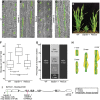
BdYDA1 is required for proper spacing of stomata. (A-D) DIC images of cleared WT (Bd21-3) (A), bdyda1-1 mutant (B), bdyda1-1 complemented with BdYDA1pro:BdYDA1-YFP:Yt transgene (C), and bdyda1-2 (D) abaxial leaf epidermis. GCs and SCs are false-colored green and yellow, respectively. Images are of the sixth leaf from base (third from main tiller) 27 days post-germination (dpg) with WT and rescued line images being cleared leaves and bdyda1-1 images epidermal peels. bdyda1-2 image shows cleared leaf from T0 regenerant. Scale bars: 40 μm. (E) Whole-plant phenotype of bdyda1-1 mutant (middle), WT (left) and rescue (right) (5 weeks post-germination). (F) Stomatal density of bdyda1-1 mutants compared with that of WT and rescued bdyda1-1 [sixth leaf from base (third from main tiller) at 27 dpg]. n=4 individuals for WT control and n=5 for rescued plants. For each sample, five different regions of the leaf were imaged and quantified. n=5 for bdyda1-1 mutants for which four different regions of the leaf were peeled, imaged and quantified. ***P<0.001; n.s., not significant (based on Kruskal–Wallis test followed by Dunn's multiple comparisons test). In boxplot, the black horizontal line indicates the median; hinges (upper and lower edges of the box) represent versions of the upper and lower quartiles; whiskers extend to the largest observation within 1.5 interquartile ranges of the box. (G) Stomatal cluster profile as percentage of clustering of quantified stomata in F (n=566 stomata for WT controls, n=729 stomata for rescue, n=835 stomata for bdyda1-1). Clusters of four or more stomata were grouped in last category ‘4+ -mer’. (H) Schematics of representative patterns of GC and SC clusters in bdyda1-1. (I) Gene/protein diagram of BdYDA1. The vertical magenta bar indicates the bdyda1-1 EMS mutation and the green bar indicates the bdyda1-2 CRISPR/Cas9-induced mutation. Model generated in Gene Structure Display Server (Hu et al., 2015).
Fig. 3.
Fig. 3.
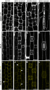
Imaging early development indicates that BdYDA1 is expressed throughout the stomatal lineage and that the initial defect in bdyda1-1 appears to be improper enforcement of non-stomatal fates. (A-H) Confocal images of progression of cells though four stages (as defined in Fig. 1A) of stomatal development in bdyda1-1 mutants (A-D) and WT (Bd21-3) (E-H) (emerging second leaf at 6 dpg, stained with PI). (I-L) Expression of rescuing BdYDA1pro:BdYDA1-YFP:Yt in bdyda1-1 (T1 plant; emerging second leaf at 6 dpg; YFP channel only). Arrowheads in I and J indicate accumulations of transgene signal. Scale bars: 10 μm. All images are oriented with the base of the leaf blade (younger cells) towards the bottom and the tip of the leaf (older cells) towards the top.
Fig. 4.
Fig. 4.
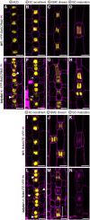
Misexpression of stomatal fate reporters in bdyda1-1 mutants is consistent with the terminal fate specification defects. Confocal images of emerging second leaf of 6 dpg T1 plants. (A-H) BdSCRM2pro:YFP-BdSCRM2 reporter in WT (Bd21-3) (A-D) and bdyda1-1 mutant (E-H) during stomatal development. Early in WT development, BdSCRM2pro:YFP-BdSCRM2 appears only in the smaller daughter of an asymmetric division (A,B). However, at the same stage in the bdyda1-1 mutant, signal is also present in mis-specified larger daughter cells (E,F). Arrowheads in E indicate examples of improper re-enforcement of non-stomatal fate in larger daughter of asymmetric division. Arrows in F point to examples of improper inhibition of division potential. (I-N) BdMUTEpro:BdMUTE-YFP reporter in WT (I-K) and bdyda1-1 mutant (L-N) during SC recruitment and GMC and SC specification. Reporter expression in WT is present only in GMCs, subsidiary mother cells (SMCs), and SCs as stomata mature. In bdyda1-1, the same reporter also marks mis-specified and clustered GMCs, SMCs and SCs. Arrowheads in L and bracket in M indicate ectopic marker expression during the SC recruitment and GMC division, respectively. Scale bars: 10 μm. Cell outlines are visualized with PI. All images are oriented with the base of the leaf (younger cells) towards the bottom and the tip of the leaf (older cells) towards the top.
Fig. 5.
Fig. 5.
bdyda1-1 mutants exhibit disruption of cell fates in other asymmetrically dividing epidermal lineages. (A-C) DIC images of cleared WT (Bd21-3) (A), bdyda1-1 mutant (B), and bdyda1-1 rescued with BdYDA1pro:BdYDA1-YFP:Yt (C) leaf epidermis. Hair cells are false-colored magenta. WT and complemented bdyda1-1 images show sixth leaf from base (third from main tiller) at 27 dpg. bdyda1-1 images show epidermal peels of sixth leaf from base (third from main tiller) at 27 dpg. Scale bars: 40 μm. (D) Hair cell density of bdyda1-1 mutants compared with that of WT and rescued bdyda1-1 [sixth leaf from base (third from main tiller) at 27 dpg]. n=4 individuals for WT control and n=5 for rescued plants. For each sample, five different regions of the leaf were imaged and quantified. n=5 for bdyda1-1 mutants for which four different regions of the leaf were peeled, imaged, and quantified. ***P<0.001; n.s., not significant (based on Kruskal–Wallis test followed by Dunn's multiple comparisons test). (E) Hair cell cluster profile as percentage of clustering of quantified hair cells in bdyda1-1 mutants, WT, and rescued bdyda1-1 (n=2286 hair cells for WT controls, n=3988 hair cells for rescue, n=2789 hair cells for bdyda1-1). Clusters of four or more hair cells were grouped in last category ‘4+ -mer’. (F-H) DIC images of cleared WT (F), bdyda1-1 mutant (G), and rescued bdyda1-1 (H) sheath epidermis. Silica cells are false-colored orange. For all genotypes, images show the sheath of the sixth leaf from base (third from main tiller) at 27 dpg. Scale bars: 40 μm. (I) Silica cell density of bdyda1-1 compared with that of WT and rescued bdyda1-1 [sheath of the sixth leaf from base (third from main tiller) at 27 dpg]. n=4 individuals for WT control, n=5 for rescued plants, and n=5 for bdyda1-1 mutants. For all, four to six different regions of the sheath were imaged and quantified. ***P<0.001; n.s., not significant (based on Kruskal–Wallis test followed by Dunn's multiple comparisons test). (J) Silica cell cluster profile as percentages of clustering of quantified silica cells in bdyda1-1 mutants, WT, and rescued bdyda1-1 (n=1328 silica cells for WT controls, n=2995 silica cells for rescue, n=1691 silica cells for bdyda1-1). Clusters of four or more silica cells were grouped in last category ‘4+ -mer’.
Fig. 6.
Fig. 6.
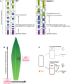
Summary of YDA's proposed role in asymmetric divisions. (A) Schematic of the bdyda1-1 phenotype and interpretations of the role of BdYDA1 in epidermal patterning of Brachypodium leaves. (B) Global and local positional information feed into developmental decisions that orient and position stomatal precursors. Global positional information in the form of lateral and longitudinal cues direct cell file identities and developmental progression of lineages, respectively. Local positional information controls fate re-enforcement to establish the correct pattern and distribution of stomata and their precursors. The relative influence of global versus local sources of positional information is likely to be species specific, i.e. longitudinally growing grass leaves are more heavily influenced by global cues and radially growing leaves with self-renewing stem-like divisions, such as in Arabidopsis, by local cues. (C) In contrast to Arabidopsis-derived pre-divisional models, which suggest that YDA mainly acts to establish physical asymmetry prior to fate establishment, we propose that YDA is primarily a post-divisional fate re-enforcer. This requires that YDA be present in both daughters of an asymmetric cell division and be available for reciprocal (and continuous) signal transduction downstream of cell-cell communication systems.

Similar articles

Cited by

References

    1. Abrash E. B. and Bergmann D. C. (2009). Asymmetric cell divisions: a view from plant development. Dev. Cell 16, 783-796. 10.1016/j.devcel.2009.05.014 - DOI - PubMed
    1. Bayer M., Nawy T., Giglione C., Galli M., Meinnel T. and Lukowitz W. (2009). Paternal control of embryonic patterning in Arabidopsis thaliana. Science 323, 1485-1488. 10.1126/science.1167784 - DOI - PubMed
    1. Bergmann D. C., Lukowitz W. and Somerville C. R. (2004). Stomatal development and pattern controlled by a MAPKK kinase. Science 304, 1494-1497. 10.1126/science.1096014 - DOI - PubMed
    1. De Smet I. and Beeckman T. (2011). Erratum: asymmetric cell division in land plants and algae: the driving force for differentiation. Nat. Rev. Mol. Cell Biol. 12, 273-273. 10.1038/nrm3094 - DOI - PubMed
    1. Dong J. and Bergmann D. C. (2010). Stomatal patterning and development. Curr. Top. Dev. Biol. 91, 267-297. 10.1016/S0070-2153(10)91009-0 - DOI - PubMed

Publication types

MeSH terms

Substances

LinkOut - more resources