Review
. 2019 Jan;44(1):166-183.
doi: 10.1038/s41386-018-0125-6. Epub 2018 Jun 19.

Sex differences in neural mechanisms mediating reward and addiction

Affiliations
Review

Sex differences in neural mechanisms mediating reward and addiction

Jill B Becker et al. Neuropsychopharmacology. 2019 Jan.

Abstract

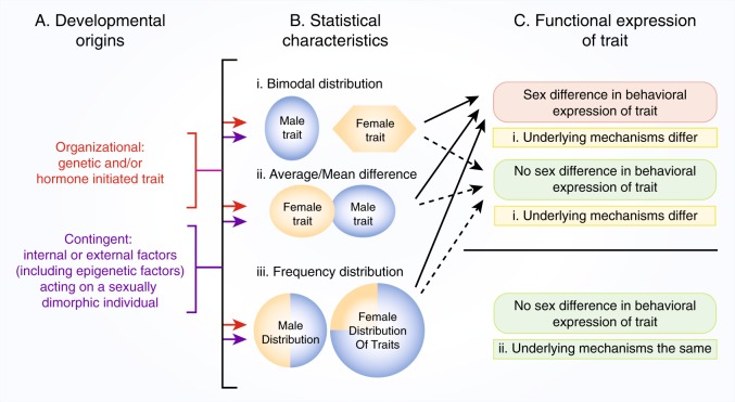
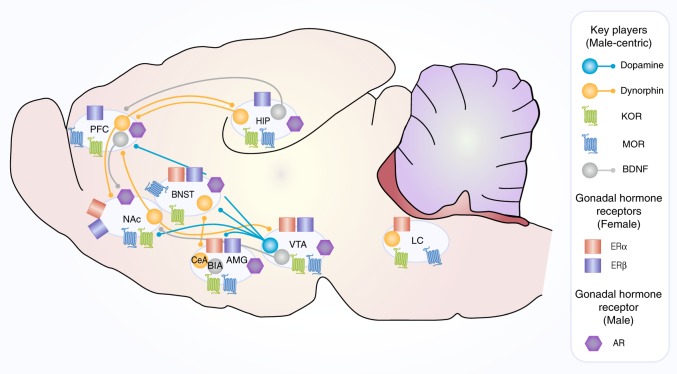
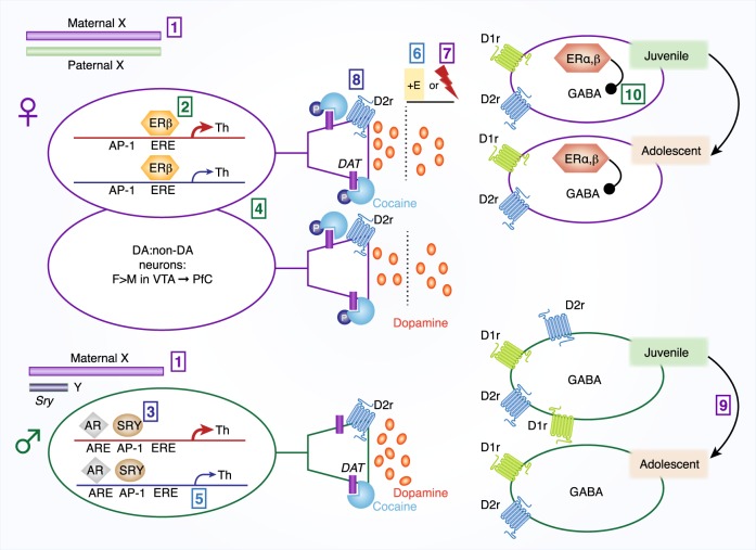
There is increasing evidence in humans and laboratory animals for biologically based sex differences in every phase of drug addiction: acute reinforcing effects, transition from occasional to compulsive use, withdrawal-associated negative affective states, craving, and relapse. There is also evidence that many qualitative aspects of the addiction phases do not differ significantly between males and females, but one sex may be more likely to exhibit a trait than the other, resulting in population differences. The conceptual framework of this review is to focus on hormonal, chromosomal, and epigenetic organizational and contingent, sex-dependent mechanisms of four neural systems that are known-primarily in males-to be key players in addiction: dopamine, mu-opioid receptors (MOR), kappa opioid receptors (KOR), and brain-derived neurotrophic factor (BDNF). We highlight data demonstrating sex differences in development, expression, and function of these neural systems as they relate-directly or indirectly-to processes of reward and addictive behavior, with a focus on psychostimulants and opioids. We identify gaps in knowledge about how these neural systems interact with sex to influence addictive behavior, emphasizing throughout that the impact of sex can be highly nuanced and male/female data should be reported regardless of the outcome.


Conflict of interest statement

The authors declare that they have no competing interests.

Figures

Fig. 1
Fig. 1
Developmental origins, statistical characteristics, and functional expression of sex differences in the brain. a Developmental origins of sex differences may arise from organizational influences or be contingent on interaction with internal or external factors. Organizational origins are defined as genetic (XY /XX chromosomes), gonadal hormone influences during critical/sensitive periods of development, and placental influences. Contingent origins include internal or external factors that include epigenetic traits induced by environmental exposure, effects of stress in utero or postnatal, nutritional factors, etc. b Statistical Characteristics that describe different types of sex differences. These occur due to multiple developmental processes. Sex differences may exist in four forms, three of which involve differences in behavioral output: i. Bimodal Distribution; ii. Average or Mean differences; iii. Frequency Distribution differences in trait occurrence. The fourth form of sex difference occurs when behavioral expression of a trait is statistically similar between males and females, but the underlying mechanisms are significantly different. c Functional Expression of Trait. Traits may be expressed differently in females and males, or the sexes may show similar expression of the trait (by the measures used) but get to the trait by different underlying mechanisms (i) or via the same mechanism (ii)
Fig. 2
Fig. 2
Synthesis of gonadal hormones. The primary gonadal hormones: progesterone, testosterone and estradiol are part of the same synthetic pathway as described in this figure. In females all steps of the synthetic pathway occur in the ovary. Since steroid hormones are not sequestered in vesicles they are secreted upon synthesis. In males, testosterone is produced by the testes. The dashed line is intended to indicate that testosterone from the testes can be converted to estradiol or dihydrotestosterone in the brain and other organs (e.g., skin) that have the necessary enzymes: aromatase or 5alpha-reductase (enzymes are depicted above enclosed in an oval). The brain is also capable of synthesizing progesterone, testosterone and estradiol de novo
Fig. 3
Fig. 3
Simplified neuroanatomical framework for major interactions between the four key players discussed in the section “Key players” (dopamine, DYN/KOR, MOR, and BDNF—data collected primarily from male rodents) and gonadal hormone receptors (data collected from females (ERα estradiol receptor alpha, ERβ estradiol receptor beta) or males (AR androgen receptor)) within neural circuitry implicated in addictive processes. Note that both males and females can produce testosterone and estradiol, although the divergent effects of these steroid hormones are mediated, in part, by sex differences in the levels and distribution of their cognate receptors [235]. Connections among brain regions utilizing the neuromodulators (dopamine, DYN, BDNF) are indicated when known [15, 184, 236, 237, 241, 242]. KOR and MOR receptor expression is indicated when known [160]. Gonadal hormone receptor expression is indicated when known [90, 91, 94, 238, 239]. AMG amygdala, AR androgen receptor, BIA basolateral nucleus of the amygdala, BDNF brain-derived neurotrophic factor, BNST bed nucleus of the stria terminalis, CeA central nucleus of the amygdala, ERα estradiol receptor alpha, ERβ estradiol receptor beta, HIP hippocampus, KOR kappa opioid receptor, LC locus coeruleus, MOR mu-opioid receptor, NAc nucleus accumbens, PFC prefrontal cortex, VTA ventral tegmental area
Fig. 4
Fig. 4
Simplified composite schematic of organizational and contingent mechanisms leading to sex differences in dopaminergic signaling within limbic brain regions. To be inclusive, the figure collapses data from ventral tegmental area → nucleus accumbens and prefrontal cortex. Known distinctions between these projections are indicated in this figure legend and in the figure. The information in this figure is based on work reported in the section “Dopamine”, which is meant to highlight research that demonstrates preclinical connections to psychostimulant and/or opioid addiction-related behaviors. [1] In embryonic mice, sex chromosome complement, but not gonadal sex, determines TH expression in midbrain dopamine neurons. Chromosomal males have greater TH expression than chromosomal females, regardless of gonadal hormones [119]. [2] ERβ is co-localized in TH-expressing neurons of the ventral tegmental area in adult males and females and is thought to stimulate TH expression in females [124]. [3] In adult males, ARs and SRY are necessary for maintenance of TH expression. SRY knockdown decreases TH expression in males, but not females [120]. [4] Females have a higher ratio of dopaminergic to non-dopaminergic neurons projecting from the ventral tegmental area to the prefrontal cortex [96]. Whether this is true for projections to the nucleus accumbens is not known. [5] There is a maternal allele bias in the Th gene in select brain regions, not including the ventral tegmental area [58]. [6] Estradiol increases dopamine release in the striatum of females only, and it requires a lack of testosterone exposure during development [87, 88]. [7] At baseline, there are higher extracellular dopamine levels in the male, compared to the female, striatum [98, 240]. However, baseline ventral tegmental area activity and phasic dopamine release is similar between the sexes in the nucleus accumbens [118]. Electrically stimulated phasic dopamine release is higher in estrous females (when estradiol levels are high) compared to males or females in low-estradiol stages [100, 101, 103, 118]. [8] Estrous-associated increases in ventral tegmental area firing and dopamine release increase phosphorylation of DAT at threonine 53 (Thr53), which increases the affinity of cocaine for DAT. This results in a greater cocaine-induced dopamine release in females during estrous [118]. [9] Males, but not females, over-produce D1r and D2r in the striatum early in development [84]. D1rs, at least, are pruned in males during adolescence via actions of microglia [86]. D1r and D2r are primarily localized to distinct populations of neurons, but for graphical simplicity in this figure, we included them on the same neurons. [10] ERα/β interact with mGluRs on neuronal membrane via caveolin to inhibit GABAergic nucleus accumbens medium spiny neurons. This decreases inhibitory input to dopamine neurons in the ventral tegmental area and allows for greater dopamine release [90, 94]. AR androgen receptor, DAT dopamine transporter, D1r dopamine D1 receptor, D2r D2 receptor, ER estrogen receptor (α or β forms indicated when known), SRY sex-determining region Y, TH tyrosine hydroxylase

References

    1. Wagner FA, Anthony JC. From first drug use to drug dependence; developmental periods of risk for dependence upon marijuana, cocaine, and alcohol. Neuropsychopharmacology. 2002;26:479–88. - PubMed
    1. Wagner FA, Anthony JC. Male-female differences in the risk of progression from first use to dependence upon cannabis, cocaine, and alcohol. Drug Alcohol Depend. 2007;86:191–8. - PubMed
    1. NSDUH NSoDUaH. Substance Abuse and Mental Health Services Administration, Results from the 2013 National Survey on Drug Use and Health: Summary of National Findings, NSDUH Series H-48, HHS Publication No. (SMA) 14-4863. Rockville, MD: Substance Abuse and Mental Health Services Administration, 2014.
    1. Cotto JH, Davis E, Dowling GJ, Elcano JC, Staton AB, Weiss SR. Gender effects on drug use, abuse, and dependence: a special analysis of results from the National Survey on Drug Use and Health. Gend Med. 2010;7:402–13. - PubMed
    1. McHugh RK, Votaw VR, Sugarman DE, Greenfield SF. Sex and gender differences in substance use disorders. Clin Psychol Rev. 2017 Nov 10. pii: S0272-7358(17)30250-7. 10.1016/j.cpr.2017.10.012. [Epub ahead of print]. PMID: 29174306. - PMC - PubMed

Publication types