Cerebral photoreception in mantis shrimp
- PMID: 29946145
- PMCID: PMC6018774
- DOI: 10.1038/s41598-018-28004-w
Cerebral photoreception in mantis shrimp
Abstract
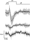
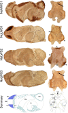
The currently unsurpassed diversity of photoreceptors found in the eyes of stomatopods, or mantis shrimps, is achieved through a variety of opsin-based visual pigments and optical filters. However, the presence of extraocular photoreceptors in these crustaceans is undescribed. Opsins have been found in extraocular tissues across animal taxa, but their functions are often unknown. Here, we show that the mantis shrimp Neogonodactylus oerstedii has functional cerebral photoreceptors, which expands the suite of mechanisms by which mantis shrimp sense light. Illumination of extraocular photoreceptors elicits behaviors akin to common arthropod escape responses, which persist in blinded individuals. The anterior central nervous system, which is illuminated when a mantis shrimp's cephalothorax protrudes from its burrow to search for predators, prey, or mates, appears to be photosensitive and to feature two types of opsin-based, potentially histaminergic photoreceptors. A pigmented ventral eye that may be capable of color discrimination extends from the cerebral ganglion, or brain, against the transparent outer carapace, and exhibits a rapid electrical response when illuminated. Additionally, opsins and histamine are expressed in several locations of the eyestalks and cerebral ganglion, where any photoresponses could contribute to shelter-seeking behaviors and other functions.
Conflict of interest statement
The authors declare no competing interests.
Figures


Similar articles
-
Opsin Expression in the Central Nervous System of the Mantis Shrimp Neogonodactylus oerstedii.Biol Bull. 2017 Aug;233(1):58-69. doi: 10.1086/694421. Epub 2017 Oct 25. Biol Bull. 2017. PMID: 29182505
-
Exceptional diversity of opsin expression patterns in Neogonodactylus oerstedii (Stomatopoda) retinas.Proc Natl Acad Sci U S A. 2020 Apr 21;117(16):8948-8957. doi: 10.1073/pnas.1917303117. Epub 2020 Apr 2. Proc Natl Acad Sci U S A. 2020. PMID: 32241889 Free PMC article.
-
Biological sunscreens tune polychromatic ultraviolet vision in mantis shrimp.Curr Biol. 2014 Jul 21;24(14):1636-1642. doi: 10.1016/j.cub.2014.05.071. Epub 2014 Jul 3. Curr Biol. 2014. PMID: 24998530
-
Parallel processing and image analysis in the eyes of mantis shrimps.Biol Bull. 2001 Apr;200(2):177-83. doi: 10.2307/1543312. Biol Bull. 2001. PMID: 11341580 Review.
-
Diverse Distributions of Extraocular Opsins in Crustaceans, Cephalopods, and Fish.Integr Comp Biol. 2016 Nov;56(5):820-833. doi: 10.1093/icb/icw022. Epub 2016 Jun 1. Integr Comp Biol. 2016. PMID: 27252200 Review.
Cited by
-
The evolution of red color vision is linked to coordinated rhodopsin tuning in lycaenid butterflies.Proc Natl Acad Sci U S A. 2021 Feb 9;118(6):e2008986118. doi: 10.1073/pnas.2008986118. Proc Natl Acad Sci U S A. 2021. PMID: 33547236 Free PMC article.
-
Exposure to Artificial Light at Night and the Consequences for Flora, Fauna, and Ecosystems.Front Neurosci. 2020 Nov 16;14:602796. doi: 10.3389/fnins.2020.602796. eCollection 2020. Front Neurosci. 2020. PMID: 33304237 Free PMC article. Review.
-
On the importance of integrating comparative anatomy and One Health perspectives in anatomy education.J Anat. 2022 Mar;240(3):429-446. doi: 10.1111/joa.13570. Epub 2021 Oct 24. J Anat. 2022. PMID: 34693516 Free PMC article. Review.
-
Light organ photosensitivity in deep-sea shrimp may suggest a novel role in counterillumination.Sci Rep. 2020 Mar 11;10(1):4485. doi: 10.1038/s41598-020-61284-9. Sci Rep. 2020. PMID: 32161283 Free PMC article.
-
Increasing complexity of opsin expression across stomatopod development.Ecol Evol. 2023 May 26;13(5):e10121. doi: 10.1002/ece3.10121. eCollection 2023 May. Ecol Evol. 2023. PMID: 37250447 Free PMC article.
References
-
- Hanna WJB, Horne JA, Renninger GH. Circadian photoreceptor organs in Limulus. II. The telson. J Comp Physiol A. 1988;162:133–140. doi: 10.1007/BF01342710. - DOI
-
- Horne JA, Renninger GH. Photoreceptor organs in Limulus. I. Ventral, median, and lateral eyes. J Comp. Physiol. A. 1988;162:127–132. doi: 10.1007/BF01342709. - DOI
-
- Wilkens LA, Larimer JL. Photosensitivity in the sixth abdominal ganglion of decapod crustaceans: a comparative study. J. Comp. Physiol. 1976;106:69–75. doi: 10.1007/BF00606572. - DOI
MeSH terms
Substances
LinkOut - more resources
Full Text Sources
Other Literature Sources

