. 2019 Jan;33(1):64-74.
doi: 10.1038/s41375-018-0180-3. Epub 2018 Jun 26.

Coexpression profile of leukemic stem cell markers for combinatorial targeted therapy in AML

Affiliations

Coexpression profile of leukemic stem cell markers for combinatorial targeted therapy in AML

S Haubner et al. Leukemia. 2019 Jan.

Abstract

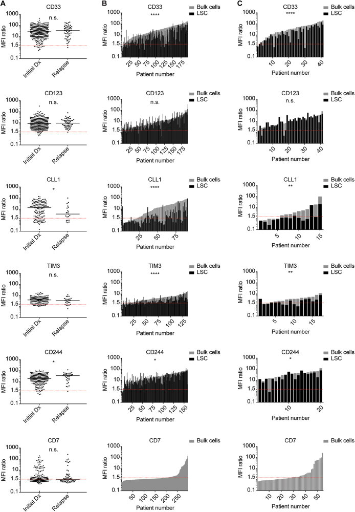
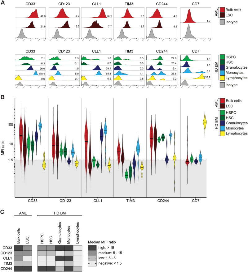
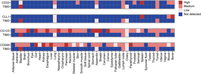
Targeted immunotherapy in acute myeloid leukemia (AML) is challenged by the lack of AML-specific target antigens and clonal heterogeneity, leading to unwanted on-target off-leukemia toxicity and risk of relapse from minor clones. We hypothesize that combinatorial targeting of AML cells can enhance therapeutic efficacy without increasing toxicity. To identify target antigen combinations specific for AML and leukemic stem cells, we generated a detailed protein expression profile based on flow cytometry of primary AML (n = 356) and normal bone marrow samples (n = 34), and a recently reported integrated normal tissue proteomic data set. We analyzed antigen expression levels of CD33, CD123, CLL1, TIM3, CD244 and CD7 on AML bulk and leukemic stem cells at initial diagnosis (n = 302) and relapse (n = 54). CD33, CD123, CLL1, TIM3 and CD244 were ubiquitously expressed on AML bulk cells at initial diagnosis and relapse, irrespective of genetic characteristics. For each analyzed target, we found additional expression in different populations of normal hematopoiesis. Analyzing the coexpression of our six targets in all dual combinations (n = 15), we found CD33/TIM3 and CLL1/TIM3 to be highly positive in AML compared with normal hematopoiesis and non-hematopoietic tissues. Our findings indicate that combinatorial targeting of CD33/TIM3 or CLL1/TIM3 may enhance therapeutic efficacy without aggravating toxicity in immunotherapy of AML.


Conflict of interest statement

The authors declare that they have no conflict of interest.

Figures

Fig. 1
Fig. 1
Antigen Expression on AML Bulk Cells and LSC at Initial Diagnosis and Relapse. Antigen expression (MFI ratio) on primary AML samples at initial diagnosis and relapse was determined via flow cytometry. Each dot or bar represents one patient sample. Red dotted line indicates MFI ratio of 1.5 as cutoff for positivity. a Initial diagnosis vs. relapse: unpaired analysis of AML bulk cells. b Initial diagnosis: paired analysis of AML bulk cells (gray) and LSC (black). c Relapse: paired analysis of AML bulk cells (gray) and LSC (black)
Fig. 2
Fig. 2
Antigen Expression in AML and Normal Hematopoiesis. Antigen expression on primary AML samples at initial diagnosis and healthy donor-derived bone marrow cell populations (HD BM) as indicated in legend. a Representative primary AML sample and healthy donor-derived bone marrow sample. Histograms indicate fluorescence intensity. Numbers indicate MFI ratio. b Antigen expression in AML and normal hematopoiesis, shown as MFI ratio. Dots indicate measured samples. Violin plots illustrate distribution of antigen expression for each analyzed cell population. Black dotted line indicates MFI ratio of 1.5 as cutoff for positivity. c Summary of antigen expression levels (median MFI ratio)
Fig. 3
Fig. 3
Antigen Coexpression in AML and Normal Hematopoiesis. Antigen coexpression on primary AML cells and on normal hematopoietic cells of healthy donor-derived bone marrow (HD BM). Each dot indicates one independent sample measurement and the MFI ratios for the respective pair of antigens. Colors indicate different cell populations. Colored areas indicate density of dots. Black lines indicate MFI ratio of 1.5 as cutoff for positivity. Coexpression rate indicates the proportion of samples with antigen coexpression (MFI ratio > 1.5 for both antigens) in relation to all measured samples. a Suitable target antigen combinations. b Unsuitable target antigen combinations. c Target antigen combinations including CD7. d Summary of antigen coexpression data. Arrows indicate suitable target antigen combinations
Fig. 4
Fig. 4
Antigen coexpression in normal tissues. Protein expression levels in normal tissues were defined by using a previously reported integrated dataset. Paired expression is illustrated for possible target antigen combinations CD33/TIM3, CLL1/TIM3, CD123/TIM3 and CD244/TIM3. Protein expression data range from 0 (not detected) to 3 (high)

References

    1. Dohner H, Estey E, Grimwade D, Amadori S, Appelbaum FR, Buchner T, et al. Diagnosis and management of AML in adults: 2017 ELN recommendations from an international expert panel. Blood. 2017;129:424–47. doi: 10.1182/blood-2016-08-733196. - DOI - PMC - PubMed
    1. Breems DA, Van Putten WL, Huijgens PC, Ossenkoppele GJ, Verhoef GE, Verdonck LF, et al. Prognostic index for adult patients with acute myeloid leukemia in first relapse. J Clin Oncol. 2005;23:1969–78. doi: 10.1200/JCO.2005.06.027. - DOI - PubMed
    1. Shlush LI, Mitchell A, Heisler L, Abelson S, Ng SWK, Trotman-Grant A, et al. Tracing the origins of relapse in acute myeloid leukaemia to stem cells. Nature. 2017;547:104–8. doi: 10.1038/nature22993. - DOI - PubMed
    1. Kantarjian H, Stein A, Gokbuget N, Fielding AK, Schuh AC, Ribera JM, et al. Blinatumomab versus chemotherapy for advanced acute lymphoblastic leukemia. N Engl J Med. 2017;376:836–47. doi: 10.1056/NEJMoa1609783. - DOI - PMC - PubMed
    1. Davila ML, Riviere I, Wang X, Bartido S, Park J, Curran K, et al. Efficacy and toxicity management of 19-28z CAR T cell therapy in B cell acute lymphoblastic leukemia. Sci Transl Med. 2014;6:224ra25. doi: 10.1126/scitranslmed.3008226. - DOI - PMC - PubMed

Publication types

MeSH terms