. 2018 Jun 12:9:683.
doi: 10.3389/fphys.2018.00683. eCollection 2018.

Assessment of the Health Status of Mussels Mytilus galloprovincialis Along the Campania Coastal Areas: A Multidisciplinary Approach

Affiliations

Assessment of the Health Status of Mussels Mytilus galloprovincialis Along the Campania Coastal Areas: A Multidisciplinary Approach

Francesca Carella et al. Front Physiol. .

Abstract

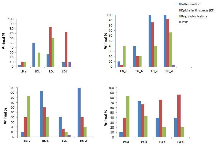
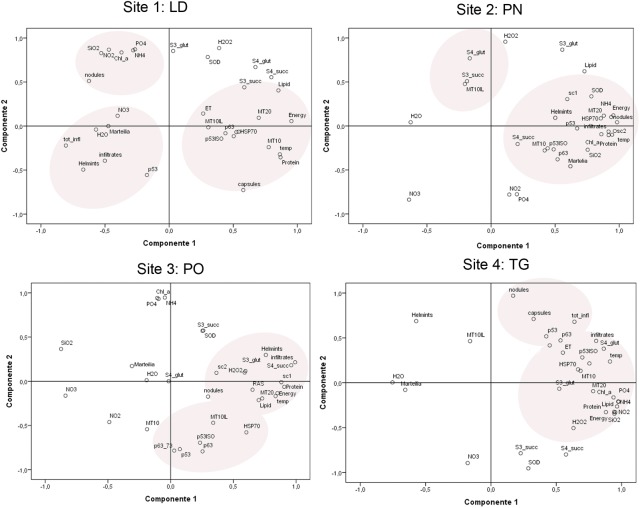
The bivalve Mytilus galloprovincialis has a broad geographic distribution, represent an important species for the ecology of coastal waters, also constituting a major aquaculture species. In the present work, molecular and tissue biomarkers were examined in mussel populations (M. galloprovincialis) located in four different areas of the coastal water of the Campania Region. During an annual life cycle, we analyzed the expression patterns of several genes commonly used to estimate cellular stress response and damage, namely p53, p63, HSP70, MT-10, and MT-20, related tissue lesions (pathogens, inflammations, digestive tubules damage), oxidative stress indicators (H2O2, SOD specific activity) and associated environmental data. The computed Principal Component Analysis showed that the areas were discernible based on the environmental data and biomarker results. About animal health status, mussels from Gulf of Pozzuoli and Naples's harbor did show a thinnest epithelial cell of digestive tubules compared to mussels sampled from other sampling sites; moreover, high prevalence of cases of intersex in three of the examinated areas were observed. The presence of a potential zoonotic pathogen (Nocardia crassostreae) was identified, appearing as an important possible emerging disease. We also reported the OIE notifiable protozoa Marteilia refringens in three areas out of four. The likely impact of both observed pathogens on the mussel health and shellfish aquaculture needs to be urgently addressed. Results are discussed considering animal histopathological health parameters and biological effects.

Keywords: biomarkers; cell damage; environmental stressors; health status; mitochondrial function; mussel.


Figures

FIGURE 1
FIGURE 1
Pathogens and diseases observed during the study: (A) Inflammatory capsule () surrounding trematodes (t) parasites at mussel foot level, (Scale bar: 100 μM); (B) unknown metacercaria at gill level (g) with no immune response from the host (Scale bar: 50 μM); (C) M. refringens (arrows) in the digestive tubules (DT) of mussels, (Scale bar: 10 μM). V.O.F. Stain; (D) nodulation at gondal level in infection by S. mytilovum (insert, arrowhead) (Scale bar: 50 μM); (E) DSD (Disorders of Sex Development) at gonadal level: note the presence of both male and female follicles in the same individual (Scale bar: 100 μM); (F) intraepithelial haemocytes (arrows) at gill level containing lipofuscin (arrows), (Scale bar: 50 μM) insert: normal gills; e, epithelia; ci, cilia; , haemocytes.
FIGURE 2
FIGURE 2
Regressive and inflammatory lesion at digestive gland level observed in the studied areas in sampled mussels M. galloprovincialis; (A) Digestive tubules (DT), showing a normal adsorbing epithelia (TE); (B) Atrophic digestive tubules (DT) with epithelia thinning (arrows) in PN; (C) Colliquative Necrosis (TN) of digestive tubules in LD; (D) Infiltrative inflammation () among digestive tubules; (E) Inflammatory nodules (arrowheads) occupying most of the digestive tissue; (F) Lipofuscin accumulation in digestive cells of DT underlined by Schmorl staining and visible as yellow pigment in H&E stain (insert). Scale bar: 50 μM. TN, tissue Necrosis; Te, tubule epithelium; DT, digestive tubules; , haemocytes.
FIGURE 3
FIGURE 3
Recorded epithelial thickness (ET) in the sampled areas (PO, PN, TG, LD). Values from 1 to 4 referred to an increased percentage of atrophic digestive tubules as reported in Kim et al. (2006).
FIGURE 4
FIGURE 4
Prevalence of the pathological conditions (inflammation, epithelial thickness-ET, Regressive lesions and Disorsers of Sex Development – DSD) observed in the different tissues in M. galloprovincialis collected at the different sites and periods.
FIGURE 5
FIGURE 5
Relative expression ratio of MT-10, MT-20 p53, p63, and HSP70 genes in the digestive gland tissue of M. galloprovincialis collected at the different sites and periods. Bars indicate the standard error.
FIGURE 6
FIGURE 6
Body composition (Water, Lipid, Protein, Energy) of M. galloprovincialis collected at the different sites and periods. Different superscripted letters indicate statistically significant differences (P < 0.05).
FIGURE 7
FIGURE 7
Hydrogen peroxide (H2O2) and SOD (Superoxide Dismutase) of M. galloprovincialis collected at the different sites and periods. Different superscripted letters indicate statistically significant differences (P < 0.05).
FIGURE 8
FIGURE 8
Mitochondrial respiration rates and oxidative stress of M. galloprovincialis collected at the different sites and periods. Different superscripted letters indicate statistically significant differences (P < 0.05).
FIGURE 9
FIGURE 9
Principal component analysis and site groupings showing the relationship between mussel biomarker responses in the considered areas.

Similar articles

Cited by

References

    1. Abele D., Philipp E., Gonzalez P., Puntarulo S. (2007). Marine invertebrate mitochondria and oxidative stress. Front. Biosci. 12 933–946. 10.2741/2115 - DOI - PubMed
    1. Aceto S., Formisano G., Carella F., De Vico G., Gaudio L. (2011). The metallothionein genes of Mytilus galloprovincialis: genomic organization, tissue expression and evolution. Mar. Genomics 4 61–68. 10.1016/j.margen.2011.01.001 - DOI - PubMed
    1. Albanese S., De Vivo B., Lima A., Cicchella D., Civitillo D., Cosenza A. (2010). Geochemical baselines and risk assessment of the Bagnoli brownfield site coastal sea sediments (Naples, Italy). J. Geochem. Explor. 105 19–33. 10.1016/j.gexplo.2010.01.007 - DOI
    1. Altavista P., Belli S., Bianchi F., Binazzi A., Comba P., Del Giudice R., et al. (2004). Cause-specific mortality in an area of Campania with numerous waste disposal sites. Epidemiol. Prev. 28 311–321. - PubMed
    1. Arienzo M., Donadio C., Mangoni O., Bolinesi F., Stanislao C., Trifuoggi M., et al. (2017). Characterization and source apportionment of PAHS in the sediments of the Gulf of Pozzuoli (Campania, Italy). Marine Poll. Bull. 124 480–487. 10.1016/j.marpolbul.2017.07.006 - DOI - PubMed