. 2018 Jun 12:9:1300.
doi: 10.3389/fimmu.2018.01300. eCollection 2018.

Resolvin E1 Promotes Bone Preservation Under Inflammatory Conditions

Affiliations

Resolvin E1 Promotes Bone Preservation Under Inflammatory Conditions

Karim El Kholy et al. Front Immunol. .

Abstract

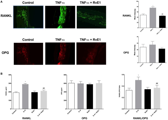
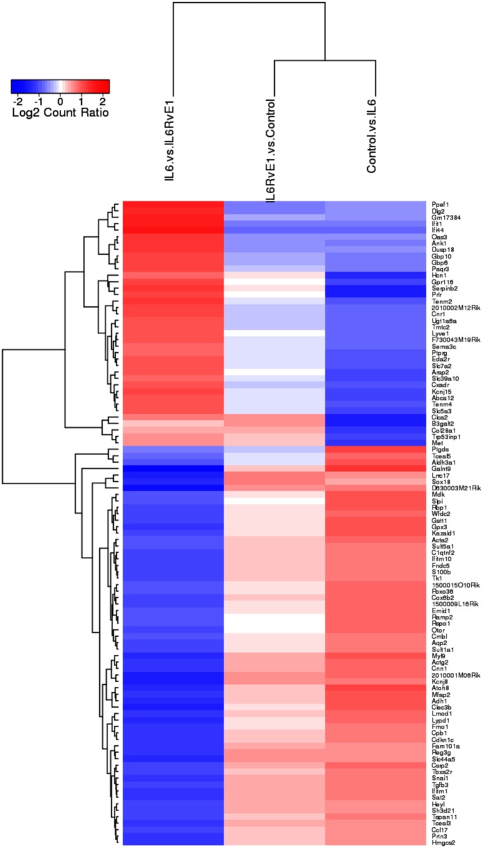
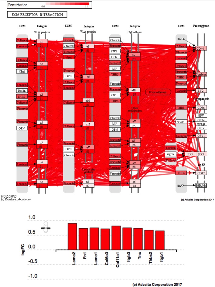
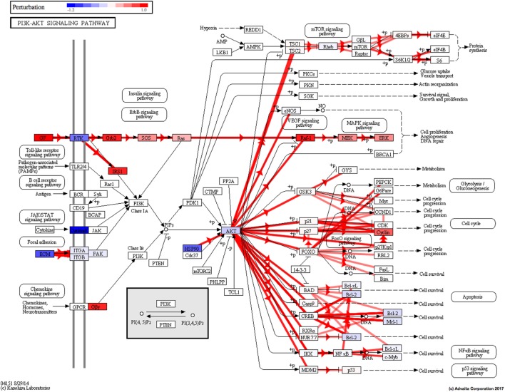
Resolvins are endogenous lipid mediators derived from omega-3 fatty acids. Resolvin E1 (RvE1), derived from eicosapentaenoic acid (EPA), modulates osteoclasts and immune cells in periodontal disease models. The direct role of RvE1 in bone remodeling is not well understood. The objective of this study was to determine the impact of RvE1 on bone remodeling under inflammatory conditions. Our working hypothesis is that RvE1 downregulates bone resorption through direct actions on both osteoblast and osteoclast function in inflammatory osteoclastogenesis. A tumor necrosis factor-α induced local calvarial osteolysis model with or without the systemic administration of RvE1 was used. To evaluate osteoclastogenesis and NFκB signaling pathway activity, murine bone tissue was evaluated by Micro CT (μCT) analysis, TRAP staining, and immunofluorescence analysis. Mechanistically, to evaluate the direct role of RvE1 impacting bone cells, primary calvarial mouse osteoblasts were stimulated with interleukin (IL)-6 (10 ng/ml) and IL-6 receptor (10 ng/ml) and simultaneously incubated with or without RvE1 (100 nM). Expression of receptor activator of NFκB ligand (RANKL) and osteoprotegerin (OPG) was measured by ELISA. RNA sequencing (RNA-Seq) and differential expression analysis was performed to determine signaling pathways impacted by RvE1. The systemic administration of RvE1 reduced calvarial bone resorption as determined by µCT. Histologic analysis of calvaria revealed that osteoclastogenesis was reduced as determined by number and size of osteoclasts in TRAP-stained sections (p < 0.05). Immunofluorescence staining of calvarial sections revealed that RvE1 reduced RANKL secretion by 25% (p < 0.05). Stimulation of osteoblasts with IL-6 increased RANKL production by 30% changing the RANKL/OPG to favor osteoclast activation and bone resorption. The ratio changes were reversed by 100 nM RvE1. RvE1 decreased the production of RANKL maintaining an RANKL/OPG more favorable for bone formation. RNA-Seq and transcriptomic pipeline analysis revealed that RvE1 significantly downregulates osteoclast differentiation mediated by differential regulation of NFκB and PI3K-AKT pathways. RvE1 reduces inflammatory bone resorption. This action is mediated, at least in part, by direct actions on bone cells promoting a favorable RANKL/OPG ratio. Mediators of resolution in innate immunity also directly regulate bone cell gene expression that is modulated by RvE1 through at least 14 specific genes in this mouse model.

Keywords: bone; bone metabolism; inflammatory diseases; resolution; resolvin E1; tissue regeneration.


Figures

Figure 1
Figure 1
(A) RvE1 reduces osteoclastogenesis triggered by TNF-α injections. TRAP-positive multinucleated giant cells (MNGCs) were counted in each section and the mean number of nuclei/cell/section was calculated by dividing the number of nuclei by the number of MNGCs found in each section. Statistically significant increases in numbers of MNGCs and nuclei/MNGC/section were measured in sections from samples receiving supracalvarial TNF-α injections when compared to samples receiving PBS injections (p < 0.05). Statistically significant decrease in both values was found in samples from animals treated with daily injections of RvE1 when compared to the TNF-α only group (p < 0.05). (B) Protective role of resolvin E1 (RvE1) in murine calvariae. Significant increases in the surface porosity (SP)/bone volume (BV) ratio was noted in specimens from the tumor necrosis factor-α (TNF-α) group when compared to the negative control and RvE1 treatment group (n = 3, p < 0.05). Reconstruction was performed using Amira 3D software. BV and SP measurements were calculated using the same software. SP/BV measurements were calculated to standardize the surface area being examined for porosity. Results are expression of the mean SP/BV ratio. *p < 0.05 vs. control. ##p < 0.05 vs. active control.
Figure 2
Figure 2
(A) Receptor activator of NFκB ligand (RANKL) and osteoprotegerin (OPG) balance is rescued by resolvin E1 (RvE1). Representative immunofluorescence imaging of primary RANKL monoclonal mouse antibodies conjugated with Alexa Fluor488. Evaluation of Immunofluorescence of histology sections in the different groups was conducted by taking 20× and 40× images and using Image J software to compare intensity of fluorescence in medullary areas. The mean immunofluorescence levels were then calculated and compared across groups. In the RvE1 treatment group, mean RANKL immunofluorescence was lower than the tumor necrosis factor-α (TNF-α) group (p < 0.05). The same slides stained with Alexa Fluor488 for RANKL immunofluorescence were also double stained with primary OPG monoclonal mouse antibodies conjugated with Dylight550 dye. Evaluation of mean OPG immunofluorescence values revealed no statistically significant differences between all three groups (p > 0.05). (B) Secreted RANKL and OPG are influenced by RvE1. The results demonstrate that RvE1 suppresses levels of RANKL via direct action on osteoblasts leading to a shift in the RANKL/OPG ratio in favor of bone preservation and formation. RvE1 at 100 nM significantly decreased RANKL production by osteoblasts stimulated by interleukin (IL)-6 (p < 0.02). Results are expressed as mean of RANKL and OPG levels. *p < 0.05 vs. control. ##p < 0.05 vs. active control.
Figure 3
Figure 3
Gene expression shift with resolvin E1 (RvE1) treatment. A heat map of top 100 differentially expressed genes (absolute log2 fold change) compares OB + interleukin (IL)-6 vs. OB alone, OB + IL-6 + RvE1 vs. OB alone, and OB + IL-6 + RvE1 vs. cells + IL-6. The color intensity reflects the level of log2 fold change for each comparison with red for upregulation and blue for downregulation. Fainter colors indicate lesser changes in gene expression between the groups being compared. Genes were ordered based on a dendrogram derived from hierarchical clustering of log2 values of all rows; genes with similar differential expression patterns were grouped together. The result demonstrates a remarkable mirror image change in gene expression. Results are expressed as mean of genes highly expressed.
Figure 4
Figure 4
Osteoclast differentiation pathway perturbation after stimulation with interleukin (IL)-6 with or without resolvin E1 (RvE1). Using Illumina’s iPathwayGuide application, a significant perturbation in the osteoclast differentiation pathway was noted after stimulation of osteoblasts with IL-6. Upregulation of IL-1 lead to promotion of downstream signaling affecting the MAPK and NFκB signaling pathways leading to upregulation of osteoclast differentiation. After treatment with RvE1, downregulation of STAT1 led to inhibition of downstream signaling affecting the MAPK and NFκB signaling pathways leading to downregulation of osteoclast differentiation.
Figure 4
Figure 4
Osteoclast differentiation pathway perturbation after stimulation with interleukin (IL)-6 with or without resolvin E1 (RvE1). Using Illumina’s iPathwayGuide application, a significant perturbation in the osteoclast differentiation pathway was noted after stimulation of osteoblasts with IL-6. Upregulation of IL-1 lead to promotion of downstream signaling affecting the MAPK and NFκB signaling pathways leading to upregulation of osteoclast differentiation. After treatment with RvE1, downregulation of STAT1 led to inhibition of downstream signaling affecting the MAPK and NFκB signaling pathways leading to downregulation of osteoclast differentiation.
Figure 5
Figure 5
Pathway impact analysis. Using Illumina’s iPathwayGuide, we identified the biological pathways with the most significant perturbation and overrepresentation combination. Extracellular matrix (ECM) receptors interactions pathway was the most significantly affected.
Figure 6
Figure 6
(A) Perturbation in extracellular matrix (ECM) receptor interactions in response to stimulation of osteoblasts with interleukin (IL)-6. Using Illumina’s iPathwayGuide, we evaluated the perturbation in ECM receptor interactions. Under inflammatory conditions, expression of ECM receptors aids in inflammatory cellular migration and adhesion. The diagram demonstrates a significant upregulation of ECM receptor interactions in response to stimulation of osteoblasts with IL-6. (B) Perturbation in ECM receptor interactions in response to resolvin E1 (RvE1) treatment after stimulation of osteoblasts with IL-6. The diagram demonstrates a significant downregulation of ECM receptor interactions in response to 100 nM RvE1 treatment after stimulation of osteoblasts with IL-6.
Figure 6
Figure 6
(A) Perturbation in extracellular matrix (ECM) receptor interactions in response to stimulation of osteoblasts with interleukin (IL)-6. Using Illumina’s iPathwayGuide, we evaluated the perturbation in ECM receptor interactions. Under inflammatory conditions, expression of ECM receptors aids in inflammatory cellular migration and adhesion. The diagram demonstrates a significant upregulation of ECM receptor interactions in response to stimulation of osteoblasts with IL-6. (B) Perturbation in ECM receptor interactions in response to resolvin E1 (RvE1) treatment after stimulation of osteoblasts with IL-6. The diagram demonstrates a significant downregulation of ECM receptor interactions in response to 100 nM RvE1 treatment after stimulation of osteoblasts with IL-6.
Figure 7
Figure 7
PI3K–AKT pathway perturbation in response to treatment of interleukin (IL)-6 stimulated osteoblasts with resolvin E1 (RvE1). The perturbation demonstrates that RvE1 influences PI3K–AKT by regulating multiple upstream and downstream targets and thereby affecting multiple cell functions including differentiation, protein synthesis, and apoptosis. These interactions also influence the NFκB, MAPK, and p53 signaling pathways.
Figure 8
Figure 8
Meta analysis of differential gene expression. We identified a group of 14, which were significantly differentiated (upregulated/downregulated) genes after induction of inflammation and were significantly reversed after treatment with resolvin E1 (RvE1). For a list of genes and their functions see Tables 1 and 2.

Similar articles

Cited by

References

    1. Thomson BM, Mundy GR, Chambers TJ. Tumor necrosis factors alpha and beta induce osteoblastic cells to stimulate osteoclastic bone resorption. J Immunol (1987) 138(3):775–9. - PubMed
    1. Thomson BM, Saklatvala J, Chambers TJ. Osteoblasts mediate interleukin 1 stimulation of bone resorption by rat osteoclasts. J Exp Med (1986) 164(1):104–12.10.1084/jem.164.1.104 - DOI - PMC - PubMed
    1. Kwan Tat S, Padrines M, Théoleyre S, Heymann D, Fortun Y. IL-6, RANKL, TNF-alpha/IL-1: interrelations in bone resorption pathophysiology. Cytokine Growth Factor Rev (2004) 15(1):49–60.10.1016/j.cytogfr.2003.10.005 - DOI - PubMed
    1. Crotti T, Smith MD, Hirsch R, Soukoulis S, Weedon H, Capone M, et al. Receptor activator NF kappaB ligand (RANKL) and osteoprotegerin (OPG) protein expression in periodontitis. J Periodontal Res (2003) 38(4):380–7.10.1034/j.1600-0765.2003.00615.x - DOI - PubMed
    1. Goldring SR. Pathogenesis of bone and cartilage destruction in rheumatoid arthritis. Rheumatology (2003) 42(Suppl 2):ii11–6.10.1093/rheumatology/keg327 - DOI - PubMed

Publication types

MeSH terms

Substances