. 2018 May 15:7:594.
doi: 10.12688/f1000research.14507.2. eCollection 2018.

A method for transplantation of human HSCs into zebrafish, to replace humanised murine transplantation models

Affiliations

A method for transplantation of human HSCs into zebrafish, to replace humanised murine transplantation models

Noémie Hamilton et al. F1000Res. .

Abstract

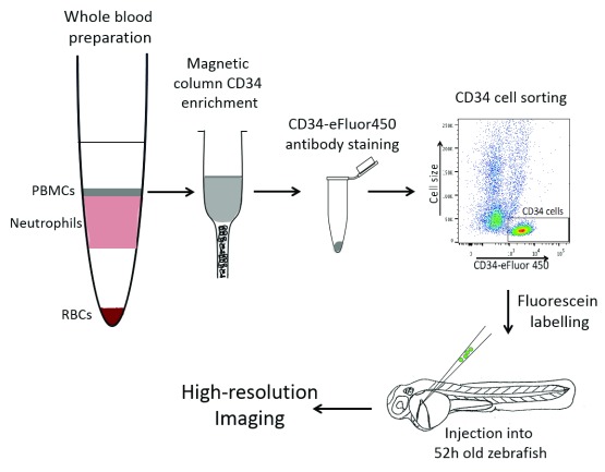
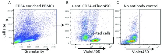
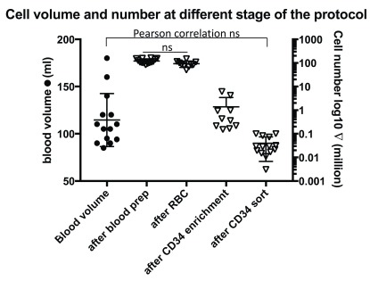
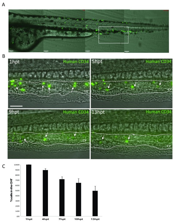
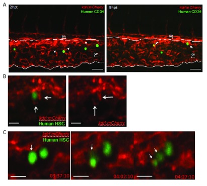
Haematopoietic stem cell (HSC) transplantation is a critical therapy for haematopoietic malignancies and immune disorders. Incomplete or delayed engraftment of HSCs in the host results in increased risk of infection and morbidity. The mechanisms of HSC engraftment are poorly understood and understanding these processes will increase transplantation success on many levels. Current animal models are immunocompromised 'humanised' mice transplanted with human HSCs. Harmful procedures include genetic manipulations and irradiation to ablate the mouse immune system, and opaque mouse tissues make visualisation of the early steps of HSC engraftment impossible. There is a need for new models to offer alternatives to humanised mice in the study of HSC transplantation. Here we described a detailed method for transplantation of human HSCs into zebrafish, before the onset of adaptive immunity. Human HSCs were purified from whole blood by enrichment of the CD34 cell population using a positive magnetic selection and further purified using an anti-CD34 antibody and cell sorting. Sorted CD34 cells were transplanted into the blood stream of 52 hour old zebrafish larvae. Human HSCs home into the zebrafish haematopoietic niche, where they engage with endothelial cells and undergo cell division. Our model offers the opportunities to image in vivo human HSC engraftment in a transparent organism, without the myeloablative strategies used in mice, and provides a unique system to understand the dynamic process of engraftment and replace current murine models. This technique can be applied to current engraftment protocols to validate the viability and efficiency of cryofrozen HSC grafts. This humanised zebrafish model will be instrumental to develop the 3Rs values in stem cell transplantation research and our detailed protocol will increase the chances of uptake of this zebrafish model by the mouse community.

Keywords: humanised zebrafish; stem cell transplantation; xenograft; zebrafish.


Conflict of interest statement

No competing interests were disclosed.

Figures

Figure 1.
Figure 1.. Diagram of our protocol: How to purify human CD34 cells from whole blood and transplant into zebrafish.
Whole blood preparation by Percoll gradient allowed us to separate peripheral blood mononuclear cells (PBMCs) from neutrophils and red blood cells (RBCs). PBMCs were enriched for CD34 cells using a positive selection magnetic column. A pure CD34 cell population was sorted using a human anti-CD34eFLuor450 antibody by FACS. CD34 cells were labelled using fluorescein and injected into the Duct of Cuvier of 52 hour post fertilisation zebrafish larvae. Animal with human cells in their CHT were selected for further high-resolution imaging.
Figure 2.
Figure 2.. Anti-CD34 antibody staining identifies a clear cell population in CD34 enriched cells by flow cytometry.
( A) Healthy cells only were gated to analyse fluorescence. ( B) A clear cell population (black rectangle) of small cells was positive for the Violet450 fluorophore as determined by the no antibody control ( C) where that same cell population is shifted to the left of the X-axis.
Figure 3.
Figure 3.. CD34 cells represent a small fraction of PBMCs.
Left scale represent the blood volume taken per donors. Paired T-test was used to analyse statistical significance between ‘after blood prep group’ and ‘after red blood cell (RBC) lysis group’ (n=10). Paired Pearson correlation analysis was performed between the blood volume taken and the final number of sorted CD34 cells and no correlation was found (p= 0.115, n=14, Pearson r=0.441).
Figure 4.
Figure 4.. Injected human CD34 cells quickly appear in the zebrafish Caudal Haematopoietic Tissue (CHT).
( A) Stitched Z-stack of whole Zebrafish larvae trunk highlighting the CHT (white rectangle). ( B) Representative Z-Stack images of fluorescein labelled human CD34 cells present at the CHT at 1hour post transplantation (hpt), 5hpt, 9hpt and 13hpt. Scale bar=80μm. ( C) Quantification of the decreasing total number of CD34 human cells in the CHT in embryos injected with fluorescein labelled human CD34 cells (n=5). First image of the CHT taken at 1hpt representing 100% of cells within the CHT and quantified following single emrbyos at 5hpt, 9hpt and 13hpt.
Figure 5.
Figure 5.. Human CD34 cells interact with zebrafish endothelial cells.
Spinning disk confocal stills of timelapses zebrafish Caudal Haematopoietic Tissue (white lines) from the endothelial cell (red) reporter line Tg(kdrl:mCherry) zebrafish larvae transplanted with human CD34 cells (green). ( A) At 2hpt, human CD34 cells are still in the vessels and empty perivascular pockets (white arrowheads) do not contain human CD34 cells. At 9hpt, human CD34 cells co-localised with endothelial cells and human CD34 cells appears inside the perivascular pockets (white arrows). DA: Dorsal aorta, CV: Caudal vein. Scale bar= 50μm. ( B) High magnification of a human CD34 cell being ‘cuddled’ by surrounding endothelial cells, note the endothelial cell protrusion acting like arms (white arrows). Single Z plane from a Z-stack. Scale bar= 50μm. N=1 ( C) Division of a human CD34 cells (white arrow) within a perivascular pocket of a 56hpf embryos, with hours post transplantation (hpt) displayed. Note the equal distribution of fluorescence between the two daughter cells in the last frame. N=1. Single Z plane from a Z-stack. Scale bar=10μm.

References

    1. Cabezas-Sainz P, Guerra-Varela J, Carreira MJ, et al. : Improving zebrafish embryo xenotransplantation conditions by increasing incubation temperature and establishing a proliferation index with ZFtool. BMC Cancer. 2018;18(1):3. 10.1186/s12885-017-3919-8 - DOI - PMC - PubMed
    1. Chi NC, Shaw RM, De Val S, et al. : Foxn4 directly regulates tbx2b expression and atrioventricular canal formation. Genes Dev. 2008;22(6):734–739. 10.1101/gad.1629408 - DOI - PMC - PubMed
    1. de Jong JL, Zon LI: Use of the zebrafish system to study primitive and definitive hematopoiesis. Annu Rev Genet. 2005;39:481–501. 10.1146/annurev.genet.39.073003.095931 - DOI - PubMed
    1. Fleming KK, Hubel A: Cryopreservation of hematopoietic and non-hematopoietic stem cells. Transfus Apher Sci. 2006;34(3):309–315. 10.1016/j.transci.2005.11.012 - DOI - PubMed
    1. Gering M, Patient R: Hedgehog signaling is required for adult blood stem cell formation in zebrafish embryos. Dev Cell. 2005;8(3):389–400. 10.1016/j.devcel.2005.01.010 - DOI - PubMed

Publication types