. 2018 Jun 12:8:185.
doi: 10.3389/fonc.2018.00185. eCollection 2018.

Expression of the Circadian Clock Gene BMAL1 Positively Correlates With Antitumor Immunity and Patient Survival in Metastatic Melanoma

Affiliations

Expression of the Circadian Clock Gene BMAL1 Positively Correlates With Antitumor Immunity and Patient Survival in Metastatic Melanoma

Leonardo Vinícius Monteiro de Assis et al. Front Oncol. .

Abstract

Introduction: Melanoma is the most lethal type of skin cancer, with increasing incidence and mortality rates worldwide. Multiple studies have demonstrated a link between cancer development/progression and circadian disruption; however, the complex role of tumor-autonomous molecular clocks remains poorly understood. With that in mind, we investigated the pathophysiological relevance of clock genes expression in metastatic melanoma.

Methods: We analyzed gene expression, somatic mutation, and clinical data from 340 metastatic melanomas from The Cancer Genome Atlas, as well as gene expression data from 234 normal skin samples from genotype-tissue expression. Findings were confirmed in independent datasets.

Results: In melanomas, the expression of most clock genes was remarkably reduced and displayed a disrupted pattern of co-expression compared to the normal skins, indicating a dysfunctional circadian clock. Importantly, we demonstrate that the expression of the clock gene aryl hydrocarbon receptor nuclear translocator-like protein 1 (BMAL1) positively correlates with patient overall survival and with the expression of T-cell activity and exhaustion markers in the tumor bulk. Accordingly, high BMAL1 expression in pretreatment samples was significantly associated with clinical benefit from immune checkpoint inhibitors. The robust intratumoral T-cell infiltration/activation observed in patients with high BMAL1 expression was associated with a decreased expression of key DNA-repair enzymes, and with an increased mutational/neoantigen load.

Conclusion: Overall, our data corroborate previous reports regarding the impact of BMAL1 expression on the cellular DNA-repair capacity and indicate that alterations in the tumor-autonomous molecular clock could influence the cellular composition of the surrounding microenvironment. Moreover, we revealed the potential of BMAL1 as a clinically relevant prognostic factor and biomarker for T-cell-based immunotherapies.

Keywords: ARNTL/BMAL1 immunotherapy; circadian rhythms; clock genes; melanoma; skin cancer.


Figures

Figure 1
Figure 1
Clinical relevance of clock genes expression in metastatic melanomas. (A) RNAseq analysis of clock genes expression in genotype-tissue expression (GTEx) normal skins (n = 234) and The Cancer Genome Atlas (TCGA) metastatic melanoma (n = 340). Expression values were estimated using RSEM and log2(x + 1) transformed. The boxes extend from the 25th to the 75th percentile, the central bold line shows the median, and whiskers are drawn from minimum to maximum values. Comparisons were performed using the two-sided Wilcoxon–Mann–Whitney test. *Significantly different from normal skin (P < 0.05). (B) Co-expression matrix showing pairwise Spearman’s correlation coefficients of clock core genes in GTEx normal skins (n = 234) and TCGA metastatic melanomas (n = 340). Networks were compared using the CoGA software. (C) Univariate Cox analysis of overall survival according to the expression of clock core genes in TCGA metastatic melanomas. Hazard Ratios including 95% confidence intervals are shown. Genes with a significant prognostic value (P < 0.05) are marked in black. (D) Kaplan–Meier survival curve according to the expression of aryl hydrocarbon receptor nuclear translocator-like protein 1 (BMAL1) in TCGA metastatic melanomas. The median expression of BMAL1 was used as the cutoff to dichotomize the population. Comparisons were performed using the log-rank test.
Figure 2
Figure 2
Aryl hydrocarbon receptor nuclear translocator-like protein 1 (BMAL1) expression positively correlates with antitumor immunity in metastatic melanomas. (A) Gene Set Enrichment Analysis (GSEA) using genes ranked according to the Spearman’s correlation coefficient between their expression and the expression of BMAL1 in The Cancer Genome Atlas (TCGA) metastatic melanomas (n = 340). Normalized enrichment scores (NES) and P-values corrected by false discovery rate (FDR) were calculated using GSEA v3.0 and Reactome pathways. Only significantly enriched pathways (adjusted P < 0.05) are shown. (B) Spearman’s correlation coefficient between the expression of BMAL1 and immune cells markers in TCGA metastatic melanomas. P-values were corrected by FDR. (C) Association between BMAL1 expression (pretreatment biopsies) and clinical benefit of melanoma patients from anti-PD1 immunotherapy (nivolumab). RNAseq data and treatment response information were obtained from Ref. (42). Comparisons were performed using the Chi-square test. CR, complete response; PR, partial response; SD, stable disease; PD, progressive disease.
Figure 3
Figure 3
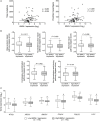
Aryl hydrocarbon receptor nuclear translocator-like protein 1 (BMAL1) expression positively correlates with the mutational and neoantigen load in metastatic melanomas. (A) Spearman’s correlation between the expression of BMAL1 and the number of total mutations and predicted neoantigens in The Cancer Genome Atlas (TCGA) metastatic melanomas (n = 68). NetMHCpan-predicted neoantigens were obtained from Ref. (40). Gene expression of (B) DNA-repair programs and (C) selected base excision repair enzymes according to the expression of BMAL1 in TCGA metastatic melanomas (n = 340). Pathway scores were calculated using single sample Gene Set Enrichment Analysis available in the GSVA R package. The median expression of BMAL1 was used as the cutoff to dichotomize the population. The boxes extend from the 25th to the 75th percentile, the central bold line shows the median, and whiskers are drawn from minimum to maximum values. Comparisons were performed using the two-sided Wilcoxon–Mann–Whitney test. *Significantly different from the low BMAL1 group.

References

    1. Garbe C, Leiter U. Melanoma epidemiology and trends. Clin Dermatol (2009) 27:3–9.10.1016/j.clindermatol.2008.09.001 - DOI - PubMed
    1. Erdei E, Torres SM. A new understanding in the epidemiology of melanoma. Expert Rev Anticancer Ther (2010) 10:1811–23.10.1586/era.10.170 - DOI - PMC - PubMed
    1. Jemal A, Siegel R, Ward E, Hao Y, Xu J, Murray T, et al. Cancer statistics, 2008. CA Cancer J Clin (2008) 58:71–96.10.3322/CA.2007.0010 - DOI - PubMed
    1. Song X, Zhao Z, Barber B, Farr AM, Ivanov B, Novich M. Overall survival in patients with metastatic melanoma. Curr Med Res Opin (2015) 31:987–91.10.1185/03007995.2015.1021904 - DOI - PubMed
    1. Chapman PB, Hauschild A, Robert C, Haanen JB, Ascierto P, Larkin J, et al. Improved survival with vemurafenib in melanoma with BRAF V600E mutation. N Engl J Med (2011) 364:2507–16.10.1056/NEJMoa1103782 - DOI - PMC - PubMed