. 2018 Oct;70(5):1389-1397.
doi: 10.1007/s10616-018-0232-6. Epub 2018 Jun 26.

Depigmenting effect of argan press-cake extract through the down-regulation of Mitf and melanogenic enzymes expression in B16 murine melanoma cells

Affiliations

Depigmenting effect of argan press-cake extract through the down-regulation of Mitf and melanogenic enzymes expression in B16 murine melanoma cells

Thouria Bourhim et al. Cytotechnology. 2018 Oct.

Abstract

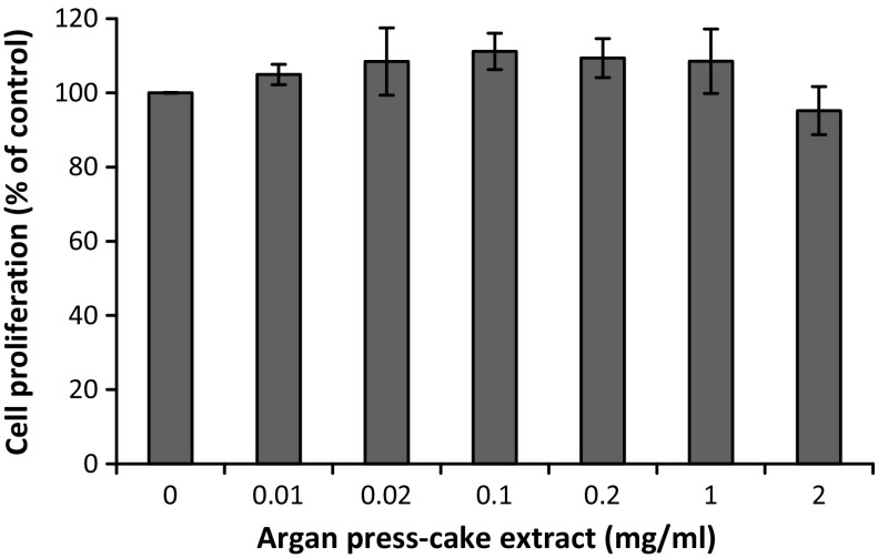
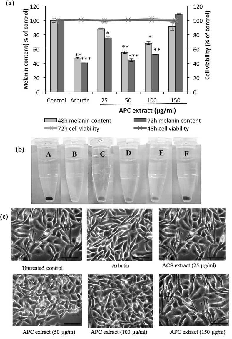
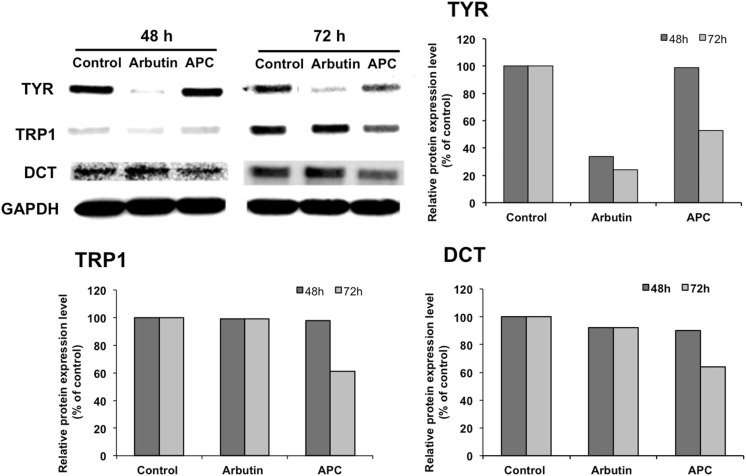
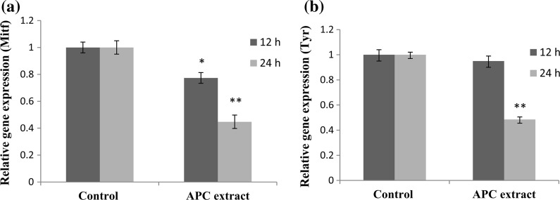
Oil extraction from the kernels of Argania spinosa (L.) Skeels (Sapotaceae), an endemic tree of Morocco, produces argan press-cake (APC) used as a shampoo and to treat sprains, scabies, and for healing wounds. We have previously reported that argan oil has antimelanogenesis effect. Here, we determined if the by-product, APC, has melanogenesis regulatory effect using B16 murine melanoma cells. The effect of APC ethanol extract on cell proliferation and melanin content of B16 cells were measured, and to elucidate the mechanism involved, the expression level of melanogenic enzymes tyrosinase (TYR), dopachrome tautomerase (DCT), and tyrosinase-related protein 1 (TRP1) were determined and mRNA expression level of microphthalmia- associated transcription factor (Mitf) and Tyr genes were quantified. APC ethanol extract showed a significant melanin biosynthesis inhibitory effect on B16 cells in a time-dependent manner without cytotoxicity, which could be due to the decreased expression of TYR, TRP1, and DCT in a time-dependent manner. APC extract down regulated Mitf and Tyr. Decreased TRP1 and DCT levels could be due to post-translational modifications. These results suggest that APC extract may be used as a new source of natural whitening products and may be introduced as an important pharmacological agent for the treatment of hyperpigmentation disorders.

Keywords: Argan press-cake; Argania spinosa; Melanogenesis; Mitf; Tyr.


Conflict of interest statement

The authors declare that they have no conflict of interest.

Figures

Fig. 1
Fig. 1
Effect of APC extract on B16 melanoma cells proliferation. After treatment of B16 cells with APC extract at various concentrations for 48 h, cell proliferation was determined by MTT assay as described in “Materials and Methods” section. Data are presented as the mean ± SD of three independent experiments. Statistical significance *P < 0.05 was defined as control versus sample
Fig. 2
Fig. 2
Effect of APC extract on the melanin content of B16 murine melanoma cells, 48 and 72 h after treatment. (a) B16 cells were seeded at a density of 5 × 105 cells per 100-mm dish. After overnight incubation, the cells were treated without (control) or with 25, 50, 100 and 150 µg/ml of APC extract for 48 and 72 h. Cells treated with 100 µmol/l arbutin served as a positive control. The synthesized melanin and total number of cells were then measured as described in “Materials and Methods” section. The melanin content was expressed as melanin content per cell and then as percentage of control (% of control). The bar graph indicates the melanin content (left-hand y-axis) while the line graph indicates cell viability (right-hand y-axis). The results are presented as the mean ± SD of three independent experiments. Statistical significance *P < 0.05 was defined as control versus sample. (b) The extracted melanin from B16 cells treated without (A) or with 100 µmol/l of arbutin (B), or with APC extract at 25 µg/ml (C), APC extract at 50 µg/ml (D), APC extract at 100 µg/ml (E), APC at 150 µg/ml (F). (c) Images of B16 cells treated with APC extract or arbutin (400× magnification)
Fig. 3
Fig. 3
Effect of APC extract on melanogenic enzymes TYR, TRP1, and DCT expression level, determined by western blot analysis (a) and their densitometric values (b). B16 melanoma cells were seeded in 100-mm dish at a density of 5 × 105 cells per dish. After overnight incubation, cells were treated without (Control) or with 50 μg/ml of APC extract and incubated for further 48 and 72 h. Total proteins were then extracted and resolved by SDS-PAGE. The resolved proteins were then blotted onto a PVDF membrane and detected by anti-TYR, anti-TRP1, anti-DCT and GAPDH antibodies (as a loading control). The signal was detected using Odyssey Fc Imaging System. Values are mean ± SD derived from three independent experiments. Statistical significance *P < 0.05 was defined as control versus sample
Fig. 4
Fig. 4
Effect of APC extract on the expression of (a) microphthalmia-associated transcription factor (Mitf) and (b) tyrosinase (Tyr) genes in B16 cells. Cells were seeded onto 100-mm dish at a density of 3 × 106 cells per dish. After overnight incubation, the cells were treated without or with APC extract (50 µg/ml) for 12 or 24 h. Real-time PCR was then used to determine the expression level of Mitf and Tyr genes. Data are expressed as the mean ± SD of three independent experiments. Statistical significance *P < 0.05 and **P < 0.01 were defined as control versus sample

References

    1. Bertolotto C, Abbe P, Hemesath TJ, Bille K, Ortonne J-P, Ballotti R. Microphthalmia gene product as a signal transducer in cAMP-induced differentiation of melanocytes. J Cell Biol. 1998;142:827–835. doi: 10.1083/jcb.142.3.827. - DOI - PMC - PubMed
    1. Byers HR, Maheshwary S, Amodeo DM, Dykstra SG. Role of cytoplasmicdynein in perinuclear aggregation of phagocytosed melanosomes and supranuclear melanin cap formation in human keratinocytes. J Investig Dermatol. 2003;121:813–820. doi: 10.1046/j.1523-1747.2003.12481.x. - DOI - PubMed
    1. Charrouf Z, Wieruszeski JM, Fkih-Tetouani S, Leroy Y, Charrouf M, Fournet B. Triterpenoid saponins from Argania spinosa. Phytochemistry. 1992;31:2079–2086. doi: 10.1016/0031-9422(92)80367-N. - DOI - PubMed
    1. El Monfalouti H, Charrouf Z, Belviso S, Ghirardello D, Scursatone B, Guillaume G, Denhez C, Zeppa G. Analysis and antioxidant capacity of the phenolic compounds from argan fruit (Argania spinosa (L.) Skeels) Eur J Lipid Sci Technol. 2012;114:446–452. doi: 10.1002/ejlt.201100209. - DOI
    1. Fitzpatrick TB, Szabo G, Seiji M, Quevedo WC. Biology of the melanin pigmentary system. In: Fitzpatrick TB, Eisen A, Wolff K, Freedberg I, Austen K, editors. Dermatology in general medicine. New York: McGraw-Hill; 1979. pp. 131–145.