

Visualization of nerve fibers around the carotid bifurcation with use of a 9.4 Tesla microscopic magnetic resonance diffusion tensor imaging with tractography
- PMID: 29947092
- PMCID: PMC6220873
- DOI: 10.1002/hed.25318
Visualization of nerve fibers around the carotid bifurcation with use of a 9.4 Tesla microscopic magnetic resonance diffusion tensor imaging with tractography
Abstract
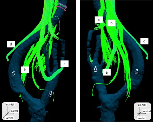
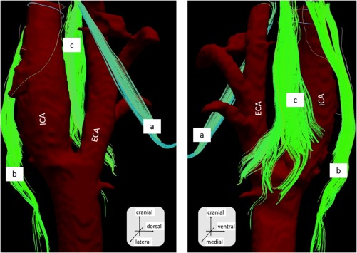
Background: Precise imaging of nerves have been challenging in the head and neck region, mainly due to low spatial resolution. Here, we investigated how nerves in the head and neck region could be visualized using an ultra-high magnetic field MR system.
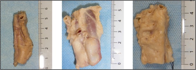
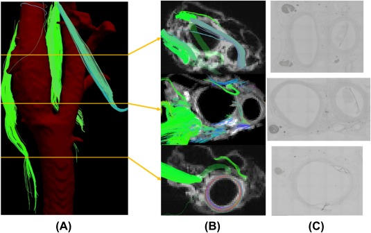
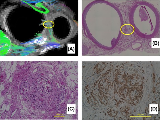
Methods: We used formol-carbol-fixed human cadaveric necks and obtained MR diffusion tensor images (DTIs) using a 9.4 Tesla (T) ultra-high magnetic field MR system. Afterward, we prepared tissue sections and checked the anatomic relationships between the neurons and the carotid artery in order to confirm that the visualized fibers are indeed neuron fibers.
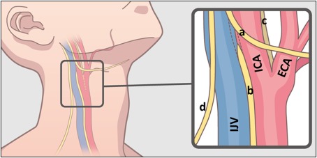
Results: We were able to identify nerves, including the vagus nerve, the hypoglossal nerve, and the spinal-accessory nerve. Hematoxylin-eosin stained histological sections confirmed neuron fibers in the same anatomic position.
Conclusion: This technique has the feasibility to be applied for a more accurate anatomic understanding, maybe even close to a histological level.
Keywords: MR diffusion tensor imaging; MR diffusion tensor tractography; head and neck; postmortem study; ultra-high magnetic field MR.
© 2018 The Authors Head & Neck Published by Wiley Periodicals, Inc.
Figures
References
-
- Overland J, Hodge JC, Breik O, Krishnan S. Surgical anatomy of the spinal accessory nerve: review of the literature and case report of a rare anatomical variant. J Laryngol Otol. 2016;130(10):969‐972. - PubMed
-
- Mori S, Zhang J. Principles of diffusion tensor imaging and its applications to basic neuroscience research. Neuron. 2006;51(5):527‐539. - PubMed
-
- Lee SP, Wu CS, Hsieh LC, Cheung WK, Chou MC. Efficacy of magnetic resonance diffusion tensor imaging and three‐dimensional fiber tractography in the detection of clinical manifestations of central nervous system lupus. Magn Reson Imaging. 2014;32(5):598‐603. - PubMed
Publication types
MeSH terms
LinkOut - more resources
Full Text Sources
Other Literature Sources
