Case Reports
. 2018 Aug;39(4):544-549.
doi: 10.1080/13816810.2018.1484929.

Two-year progression analysis of RPE65 autosomal dominant retinitis pigmentosa

Affiliations
Case Reports

Two-year progression analysis of RPE65 autosomal dominant retinitis pigmentosa

Ruben Jauregui et al. Ophthalmic Genet. 2018 Aug.

Abstract

Background: The RPE65 gene was recently described to cause autosomal dominant retinitis pigmentosa (adRP), presenting with a phenotype resembling choroideremia. This study presents the 2-year progression of RPE65 adRP in a patient.

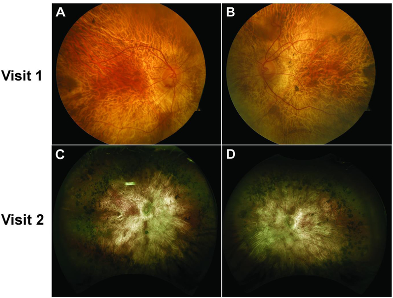
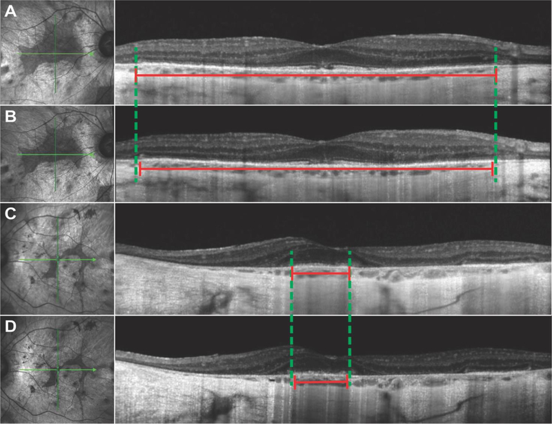
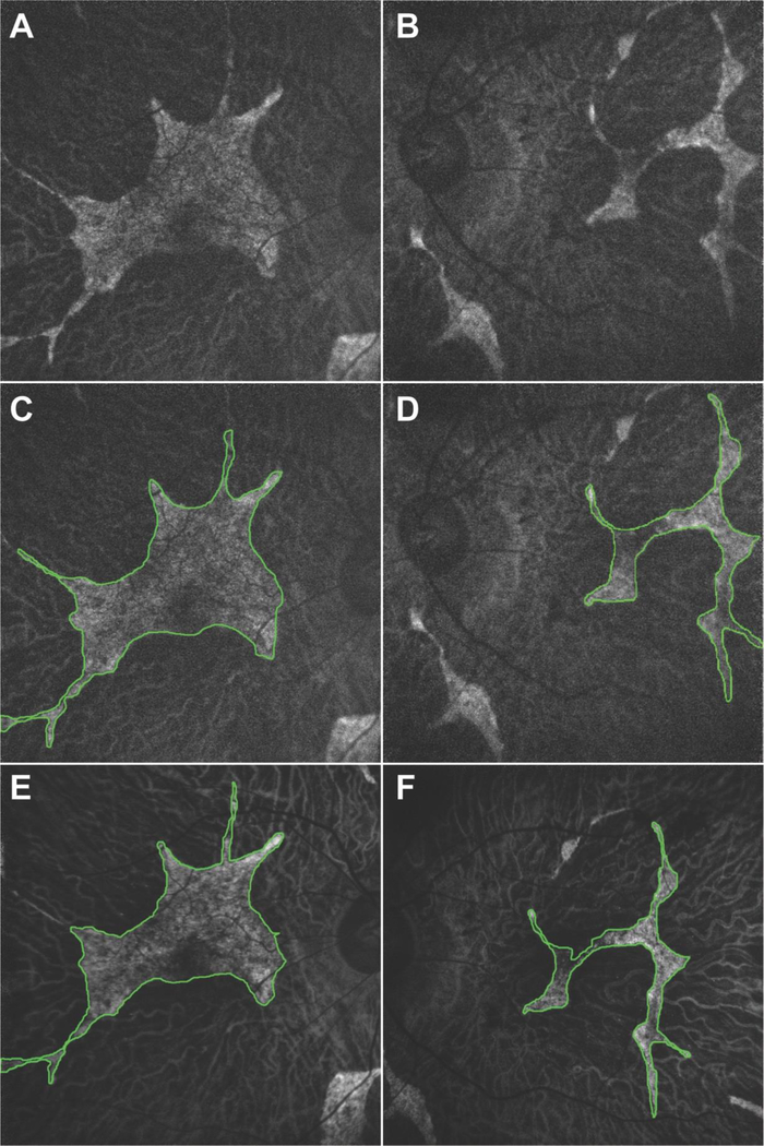
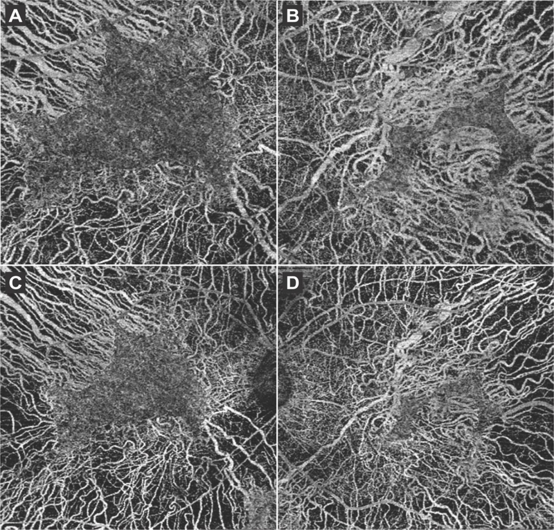
Methods: This is an observational case report of one patient. The patient received a full ophthalmic examination during both visits, including diagnostic imaging such as spectral domain optical coherence tomography (SD-OCT), OCT-angiography (OCT-A), short-wave fundus autofluorescence (FAF), and fundus photography. Genetic characterization was obtained by DNA sequencing from peripheral blood lymphocytes obtained during the first visit.

Results: RPE65 adRP phenocopied choroideremia at the initial fundoscopy. Upon the patient's return to our clinic 2 years later, DNA sequencing revealed a heterozygous mutation in the RPE65 gene. Diagnostic imaging by SD-OCT and FAF suggested disease progression. In conjunction with clinical examination and imaging, the diagnosis was revised to adRP caused by RPE65.

Conclusion: adRP due to a mutation in the gene encoding RPE65 phenocopied choroideremia. Based on our analysis of the 2-year disease progression in this patient, RPE65 adRP is mild and has a slow rate of disease progression.

Keywords: Autosomal dominant; RPE65; disease progression; retinitis pigmentosa.


Conflict of interest statement

Conflicts of interest: The authors declare that they have no conflicts of interest.

DECLARATION OF INTEREST STATEMENT

The authors report no conflicts of interest.

Figures

Figure 1.
Figure 1.
Color fundus (top row) and widefield retina photography (bottom row) images showing widespread areas of chorioretinal atrophy and exposed underlying large choroidal vessels on both the right (A, C) and left (B, D) eye at visits 1 and 2. An island of spared retina on the parafoveal region was also observed bilaterally at both visits. Extensive intraretinal pigment migration in the periphery can be seen clearly in the widefield fundus images (C, D). Despite the different imaging modalities used, no significant progression is observed between the first and second visits. The difference in color and exposure is attributed to the different imaging modalities, but an island of spared retina of similar shape and size can be appreciated on both eyes at both visits.
Figure 2.
Figure 2.
SD-OCT showed widespread peripheral retinal atrophy with disruption of the outer nuclear layer and ellipsoid zone (EZ) in both the right (A, B) and left (C, D) eye. The double-headed red lines indicate the length of the EZ line, while the dashed green lines outline the borders of the EZ line according to the first visit. A small decrease in EZ line length (2% in the right, 9% in the left eye) was measured in the second visit (B, D) as compared to the first visit (A, C).
Figure 3.
Figure 3.
Fundus autofluorescence imaging of the right (A, C, E) and left (B, D, F) eye showed generalized hypoautofluorescent areas corresponding to RPE atrophy bound by sharply demarcated borders. Areas of preserved retinal tissue were also detected on the parafoveal areas of both eyes (outlined in green). The area of preserved retinal tissue decreased in size from the first visit (A, B, C, D) to the second (E, F) visit by 17% in the right and 21% in the left eye.
Figure 4.
Figure 4.
6×6 mm OCT-A scans obtained in the first visit of both the right (A) and left (B) eye showed the underlying larger choroidal vessels in areas where the overlying retinal tissue and choriocapillaries (CC) had atrophied, whereas the CC vasculature appeared normal in spared retinal tissue. 8×8 mm OCT-A scans performed during the second visit (C, D) showed similar findings.

References

    1. Hartong DT, Berson EL, Dryja TP. Retinitis pigmentosa. Lancet 2006;368:1795–809. - PubMed
    1. Morimura H, Fishman GA, Grover SA, Fulton AB, Berson EL, Dryja TP. Mutations in the RPE65 gene in patients with autosomal recessive retinitis pigmentosa or leber congenital amaurosis. Proc Natl Acad Sci U S A 1998;95:3088–93. - PMC - PubMed
    1. Shin Y, Moiseyev G, Chakraborty D, Ma JX. A Dominant Mutation in Rpe65, D477G, Delays Dark Adaptation and Disturbs the Visual Cycle in the Mutant Knock-In Mice. Am J Pathol 2017;187:517–527. - PMC - PubMed
    1. Bowne SJ, Humphries MM, Sullivan LS, et al. A dominant mutation in RPE65 identified by whole-exome sequencing causes retinitis pigmentosa with choroidal involvement. Eur J Hum Genet 2011;19:1074–81. - PMC - PubMed
    1. Hull S, Mukherjee R, Holder GE, Moore AT, Webster AR. The clinical features of retinal disease due to a dominant mutation in RPE65. Mol Vis 2016;22:626–35. - PMC - PubMed

Publication types

Substances