. 2018 Sep 1;120(3):1356-1373.
doi: 10.1152/jn.00263.2018. Epub 2018 Jun 27.

Estrous cycle-induced sex differences in medium spiny neuron excitatory synaptic transmission and intrinsic excitability in adult rat nucleus accumbens core

Affiliations

Estrous cycle-induced sex differences in medium spiny neuron excitatory synaptic transmission and intrinsic excitability in adult rat nucleus accumbens core

Stephanie B Proaño et al. J Neurophysiol. .

Abstract

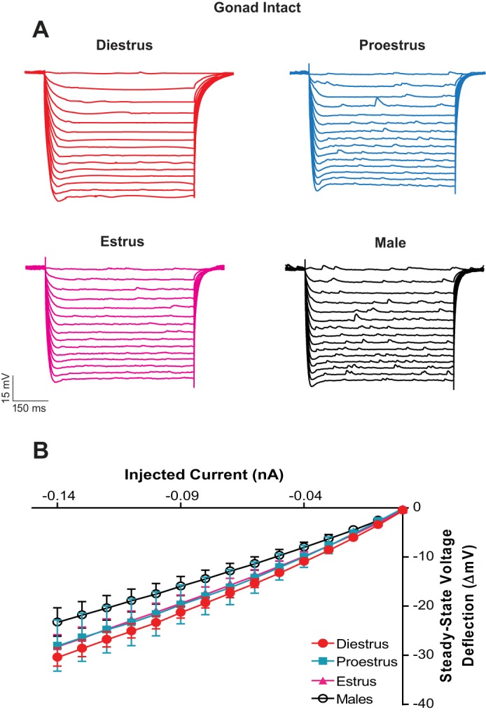
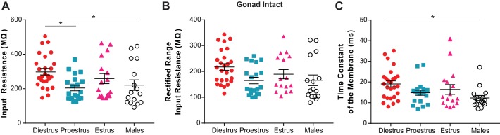
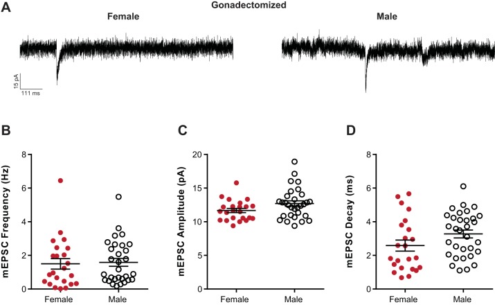
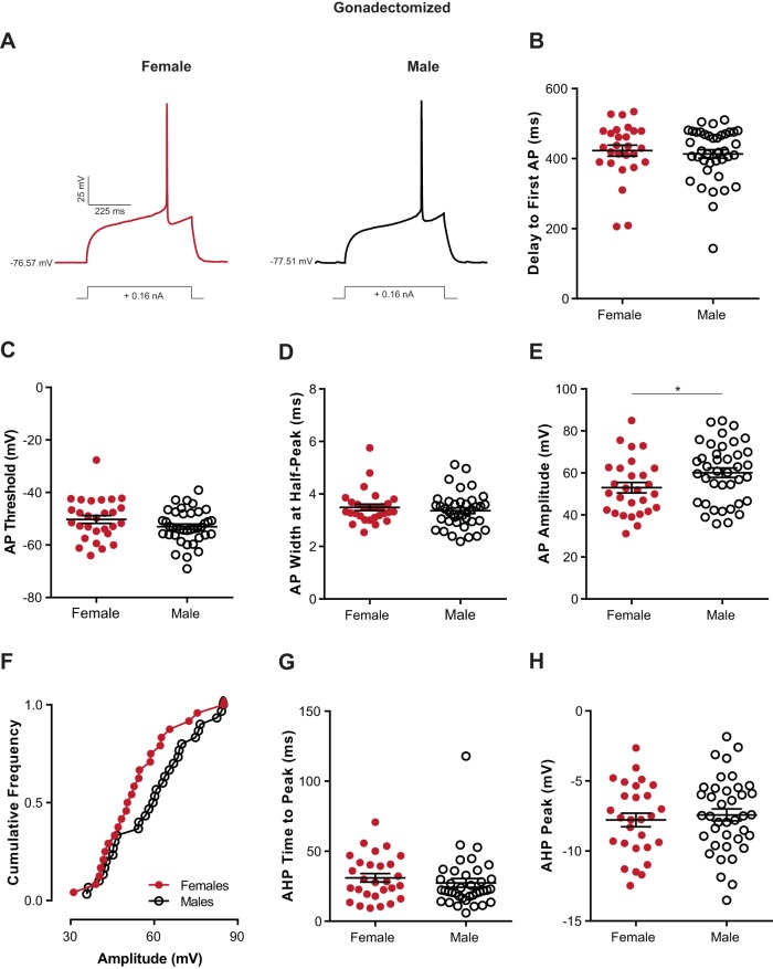
Naturally occurring hormone cycles in adult female humans and rodents create a dynamic neuroendocrine environment. These cycles include the menstrual cycle in humans and its counterpart in rodents, the estrous cycle. These hormone fluctuations induce sex differences in the phenotypes of many behaviors, including those related to motivation, and associated disorders such as depression and addiction. This suggests that the neural substrate instrumental for these behaviors, including the nucleus accumbens core (AcbC), likewise differs between estrous cycle phases. It is unknown whether the electrophysiological properties of AcbC output neurons, medium spiny neurons (MSNs), change between estrous cycle phases. This is a critical knowledge gap given that MSN electrophysiological properties are instrumental for determining AcbC output to efferent targets. Here we test whether the intrinsic electrophysiological properties of adult rat AcbC MSNs differ across female estrous cycle phases and from males. We recorded MSNs with whole cell patch-clamp technique in two experiments, the first using gonad-intact adult males and females in differing phases of the estrous cycle and the second using gonadectomized males and females in which the estrous cycle was eliminated. MSN intrinsic electrophysiological and excitatory synaptic input properties robustly changed between female estrous cycle phases and males. Sex differences in MSN electrophysiology disappeared when the estrous cycle was eliminated. These novel findings indicate that AcbC MSN electrophysiological properties change across the estrous cycle, providing a new framework for understanding how biological sex and hormone cyclicity regulate motivated behaviors and other AcbC functions and disorders. NEW & NOTEWORTHY This research is the first demonstration that medium spiny neuron electrophysiological properties change across adult female hormone cycle phases in any striatal region. This influence of estrous cycle engenders sex differences in electrophysiological properties that are eliminated by gonadectomy. Broadly, these findings indicate that adult female hormone cycles are an important factor for neurophysiology.

Keywords: estrous cycle; excitability; medium spiny neurons; nucleus accumbens; sex steroid hormones.


Figures

Fig. 1.
Fig. 1.
Location of whole cell patch-clamped medium spiny neurons (MSNs) in rat nucleus accumbens (Acb) core. A: gonad-intact females in differing estrous cycle phases and males. B: gonadectomized females and males. AC, anterior commissure; LV, lateral ventricle.
Fig. 2.
Fig. 2.
Medium spiny neuron (MSN) miniature excitatory postsynaptic current (mEPSC) properties: gonad-intact females and males. A: representative examples of mEPSCs recorded in diestrus, proestrus, and estrus female and male MSNs. B–D: mEPSC frequency (B), mEPSC amplitude (C), and mEPSC decay (D) varied across estrous cycle phases in females and/or from males. E: mEPSC amplitude was significantly correlated with mEPSC decay in diestrus, proestrus, and estrus females but not males. Horizontal line superimposed upon scatterplots in B–D indicates the mean. Lines situated above scatterplots indicate statistical significance. *P < 0.05, **P < 0.01, ***P < 0.001, ****P < 0.0001. Complete statistical information is in Table 2.
Fig. 3.
Fig. 3.
Medium spiny neuron (MSN) individual action potential properties: gonad-intact females and males. A: voltage responses of diestrus, proestrus, and estrus female and male MSNs to a single depolarizing current injection. B: delay to first action potential varies across estrous cycle and/or vs. males but remained a statistical trend. C: action potential width measured at half peak amplitude varied across estrous cycle and/or vs. males. D–G: action potential threshold (D), action potential amplitude (E), action potential afterhyperpolarization time to peak amplitude (F), and action potential afterhyperpolarization peak amplitude (G) did not vary across estrous cycle phases in females and/or vs. males. Horizontal line superimposed upon scatterplots in B–G indicates the mean. Lines situated above scatterplots indicate statistical significance. *P < 0.05. Complete statistical information is in Table 3. AP, action potential; AHP, afterhyperpolarization.
Fig. 4.
Fig. 4.
Medium spiny neuron (MSN) action potential initiation and generation: gonad-intact females and males. A: voltage responses of diestrus, proestrus, and estrus female and male MSNs to a series of depolarizing current injections. B: action potential firing rates evoked by depolarizing current injections. C and D: rheobase (C) and resting membrane potential (D) varied across estrous cycle phase in females and/or vs. males: E and F: slopes of the evoked firing rate-to-positive current curve (FI slope) in individual MSNs (E) and minimum evoked firing rate (F) did not vary across estrous cycle and/or vs. males. Horizontal line superimposed upon scatterplots in C–F indicates the mean. Lines situated above scatterplots indicate statistical significance. *P < 0.05, ***P < 0.001. Complete statistical information is in Table 3.
Fig. 5.
Fig. 5.
Medium spiny neuron (MSN) passive electrophysiological properties: gonad-intact females and males. A: voltage response of diestrus, proestrus, and estrus female and male MSNs to a series of hyperpolarizing current injections. B: the injected current-to-steady-stage voltage deflection curve (IV curve) varied across estrous cycle in females and/or vs. males.
Fig. 6.
Fig. 6.
Medium spiny neuron (MSN) input resistance and time constant of the membrane properties: gonad-intact females and males. Input resistance in the linear range (A), input resistance in the rectified range (B; overall ANOVA for input resistance in rectified range was significant, but no differences were detected by post hoc test), and time constant of the membrane (C) varied across estrous cycle in females and/or vs. males. Horizontal line superimposed upon scatterplots in A–C indicates the mean. Lines situated above scatterplots indicate statistical significance. *P < 0.05. Complete statistical information is in Table 3.
Fig. 7.
Fig. 7.
Medium spiny neuron (MSN) miniature excitatory postsynaptic current (mEPSC) properties: gonadectomized females and males. A: representative examples of mEPSCs recorded in gonadectomized female and male MSNs. B–D: mEPSC frequency (B), mEPSC amplitude (C), and mEPSC decay (D) did not vary between gonadectomized females and males. Horizontal line superimposed upon scatterplots in B–D indicates the mean. Complete statistical information is in Table 4.
Fig. 8.
Fig. 8.
Medium spiny neuron (MSN) individual action potential properties: gonadectomized females and males. A: voltage responses of gonadectomized female and male MSNs to a single depolarizing current injection. B–D: delay to first action potential (B), action potential threshold (C), and action potential width measured at half peak amplitude (D) did not vary between gonadectomized females and males: E and F: action potential amplitude appeared to vary between gonadectomized females and males (E); however, a cumulative frequency plot of action potential amplitude data distributions (F) failed to reach significance, indicating that action potential amplitude is not a robust sex difference. G and H: action potential afterhyperpolarization time to peak amplitude (G) and action potential afterhyperpolarization peak amplitude (H) did not vary between gonadectomized females and males. Horizontal line superimposed upon scatterplots in BE, G, and H indicates the mean. Lines situated above scatterplots indicate statistical significance. *P < 0.05. Complete statistical information is in Table 5. AP, action potential; AHP, afterhyperpolarization.
Fig. 9.
Fig. 9.
Medium spiny neuron (MSN) action potential initiation and generation: gonadectomized females and males. A: voltage responses of gonadectomized female and male MSNs to a series of depolarizing current injections. B: action potential firing rates evoked by depolarizing current injections. C–E: rheobase (C), slopes of the evoked firing rate-to-positive current curve (FI slope) in individual MSNs (D), and minimum firing rate (E) did not vary between gonadectomized females and males. Horizontal line superimposed upon scatterplots in C–E indicates the mean. Complete statistical information is in Table 5.
Fig. 10.
Fig. 10.
Medium spiny neuron (MSN) passive electrophysiological properties: gonadectomized females and males. A: voltage response of gonadectomized female and male MSNs to a series of hyperpolarizing current injections. B: the injected current-to-steady-stage voltage deflection curve (IV curve) did not vary between gonadectomized females and vs. males.
Fig. 11.
Fig. 11.
Medium spiny neuron (MSN) input resistance and time constant of the membrane properties: gonadectomized females and males. Input resistance in the linear range (A), input resistance in the rectified range (B), and time constant of the membrane (C) did not vary between gonadectomized females and males. Horizontal line superimposed upon scatterplots in A–C indicates the mean. Complete statistical information is in Table 5.
Fig. 12.
Fig. 12.
Schematic of nucleus accumbens core medium spiny neuron excitatory synaptic input and intrinsic excitability changes between estrous cycle phases in the adult female rat. Excitatory synaptic input properties are decreased in the diestrus phase of the cycle while intrinsic excitability is increased, as measured by changes in rheobase, resting membrane potential, and input resistance. In the proestrus and estrus phases of the cycle, excitatory synaptic input is augmented, potentially through both an increase in excitatory synapse number and other presynaptic modifications and postsynaptic AMPA receptor number and/or other properties. In contrast, intrinsic excitability is decreased. LH, luteinizing hormone; FSH, follicle-stimulating hormone.

Similar articles

Cited by

References

    1. Almey A, Milner TA, Brake WG. Estrogen receptors in the central nervous system and their implication for dopamine-dependent cognition in females. Horm Behav 74: 125–138, 2015. doi:10.1016/j.yhbeh.2015.06.010. - DOI - PMC - PubMed
    1. Alreja M. Electrophysiology of kisspeptin neurons. Adv Exp Med Biol 784: 349–362, 2013. doi:10.1007/978-1-4614-6199-9_16. - DOI - PubMed
    1. Altemus M, Sarvaiya N, Neill Epperson C. Sex differences in anxiety and depression clinical perspectives. Front Neuroendocrinol 35: 320–330, 2014. doi:10.1016/j.yfrne.2014.05.004. - DOI - PMC - PubMed
    1. Andersen SL, Thompson AP, Krenzel E, Teicher MH. Pubertal changes in gonadal hormones do not underlie adolescent dopamine receptor overproduction. Psychoneuroendocrinology 27: 683–691, 2002. doi:10.1016/S0306-4530(01)00069-5. - DOI - PubMed
    1. Arnauld E, Dufy B, Pestre M, Vincent JD. Effects of estrogens on the responses of caudate neurons to microiontophoretically applied dopamine. Neurosci Lett 21: 325–331, 1981. doi:10.1016/0304-3940(81)90225-1. - DOI - PubMed

Publication types

LinkOut - more resources