. 2018 Sep 1;120(3):1374-1385.
doi: 10.1152/jn.00158.2018. Epub 2018 Jun 27.

Adult mouse sensory neurons on microelectrode arrays exhibit increased spontaneous and stimulus-evoked activity in the presence of interleukin-6

Affiliations

Adult mouse sensory neurons on microelectrode arrays exhibit increased spontaneous and stimulus-evoked activity in the presence of interleukin-6

Bryan J Black et al. J Neurophysiol. .

Abstract

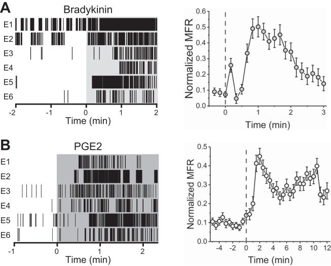
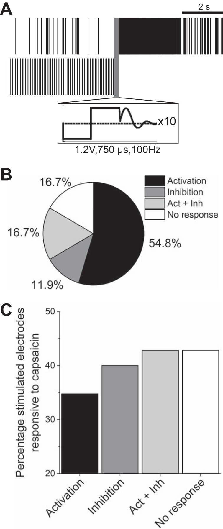
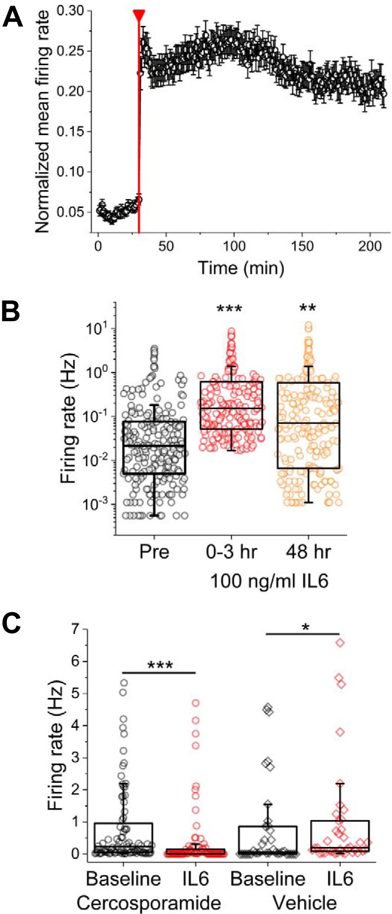
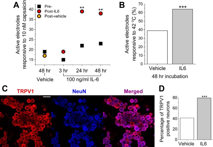
Following inflammation or injury, sensory neurons located in the dorsal root ganglia (DRG) may exhibit increased spontaneous and/or stimulus-evoked activity, contributing to chronic pain. Current treatment options for peripherally mediated chronic pain are highly limited, driving the development of cell- or tissue-based phenotypic (function-based) screening assays for peripheral analgesic and mechanistic lead discovery. Extant assays are often limited by throughput, content, use of tumorigenic cell lines, or tissue sources from immature developmental stages (i.e., embryonic or postnatal). Here, we describe a protocol for culturing adult mouse DRG neurons on substrate-integrated multiwell microelectrode arrays (MEAs). This approach enables multiplexed measurements of spontaneous as well as stimulus-evoked extracellular action potentials from large populations of cells. The DRG cultures exhibit stable spontaneous activity from 9 to 21 days in vitro. Activity is readily evoked by known chemical and physical agonists of sensory neuron activity such as capsaicin, bradykinin, PGE2, heat, and electrical field stimulation. Most importantly, we demonstrate that both spontaneous and stimulus-evoked activity may be potentiated by incubation with the inflammatory cytokine interleukin-6 (IL-6). Acute responsiveness to IL-6 is inhibited by treatment with a MAPK-interacting kinase 1/2 inhibitor, cercosporamide. In total, these findings suggest that adult mouse DRG neurons on multiwell MEAs are applicable to ongoing efforts to discover peripheral analgesic and their mechanisms of action. NEW & NOTEWORTHY This work describes methodologies for culturing spontaneously active adult mouse dorsal root ganglia (DRG) sensory neurons on microelectrode arrays. We characterize spontaneous and stimulus-evoked adult DRG activity over durations consistent with pharmacological interventions. Furthermore, persistent hyperexcitability could be induced by incubation with inflammatory cytokine IL-6 and attenuated with cercosporamide, an inhibitor of the IL-6 sensitization pathway. This constitutes a more physiologically relevant, moderate-throughput in vitro model for peripheral analgesic screening as well as mechanistic lead discovery.

Keywords: dorsal root ganglion; microelectrode arrays; nociceptors; sensitization.


Figures

Fig. 1.
Fig. 1.
Immunocytochemistry and size-based discrimination of neuronal subtypes. A and B: CGRP-positive cells (blue) indicate peptidergic C-fiber neurons (C Pep). Isolectin B4 (IB4)-positive (green), Neurofilament 200 (NF200)-negative cells indicate nonpeptidergic C-fiber neurons (C Non-pep). IB4-positive, NF200-positive (red) cells indicate Aδ-fiber neurons. NF200 (only)-positive cells indicate Aβ-fiber neurons. Scale bar represents 200 µm. C: automated measurements of cross-sectional area suggest a large percentage of small- and medium-gauge neurons, indicating a primarily nociceptive cell population.
Fig. 2.
Fig. 2.
Dorsal root ganglia (DRG) neurons exhibit well-resolved, stable spontaneous activity in vitro. A: phase image of disorganized dissociated DRG neuron culture. Scale bar represents 100 µm. B: heat map of 12-well multiwell microelectrode array recording. Each box indicates a single well. Each spot indicates electrode activity integrated over a 1-s period. C: representative trace of filtered continuous recording from a single electrode. Scale bars represent 1 s (horizontal) and 75 µV (vertical). D: representative mean waveform ± SD for a single unit. E: percentages of spontaneously active channels in terms of spontaneous firing rates. F: mean firing rate and active electrode yield over 21 days in vitro.
Fig. 3.
Fig. 3.
Spontaneously active DRG neurons exhibit various firing patterns in vitro. A: representative raster plots from 4 spontaneously active electrodes (E) within a single well exhibiting different firing patterns. B: associated interspike interval histograms.
Fig. 4.
Fig. 4.
Capsaicin and temperature responsiveness. A: representative continuous data traces from electrodes within a single well exhibiting responsiveness to 100 nM capsaicin. B: representative raster plots from 6 10 nM capsaicin-responsive electrodes (E) within a single well (left) and capsaicin concentration responsiveness as a percentage of active electrodes (right). C: representative raster plots from 6 temperature-responsive electrodes within a single well (left). Normalized mean firing rate (MFR) during calibrated temperature increase from 37 to 42°C.
Fig. 5.
Fig. 5.
Bradykinin and PGE2 responsiveness. A: representative raster plots from 6 10 nM bradykinin-responsive electrodes (E) within a single well (left) and normalized mean firing rates (MFR) before and after bradykinin addition (right). B: representative raster plots from 6 PGE2-responsive electrodes within a single well (left) and normalized mean firing rate before and after addition of PGE2.
Fig. 6.
Fig. 6.
Electrical stimulation at frequencies >100 Hz elicits differential responsiveness. A: representative raster plots from 2 stimulated electrodes exhibiting either activation (top) or inhibition (bottom) of activity. Scale bar represents 2 s. Inset (black square) illustrates cathodic-leading biphasic waveform and artifact removal. B: percentage breakdown of electrodes that were activated, inhibited, both (Act + Inh), or neither (n = 42). C: percentage of stimulated electrodes found responsive to 1 μM capsaicin.
Fig. 7.
Fig. 7.
Spontaneous sensory neuron activity is significantly increased with short- and long-term incubation with IL-6. A: normalized mean firing rate for electrode subset found to be responsive to IL-6 (41% of previously active electrodes). Red line indicates addition of 100 ng/ml IL-6. B: firing rates before (Pre), immediately following (0–3 h), and 48 h following IL-6 addition. Symbols represent data from single electrodes. C: firing rates before and immediately following IL-6 addition in the presence of vehicle (water) or MAPK-interacting kinase 1/2 inhibitor cercosporamide. *P < 0.05, **P < 0.01, and ***P < 0.001.
Fig. 8.
Fig. 8.
Incubation with IL-6 causes increased capsaicin and temperature responsiveness. A: active electrodes responsive to capsaicin before treatment (Pre-) and following 3-, 24-, or 48-h incubation with IL-6 or vehicle. All treatment recordings (Post-) were carried out 48 h after baseline. B: active electrodes responsive to 42°C following 48-h incubation with IL-6 or vehicle. C: representative fluorescence images of immunocytochemistry staining for capsaicin- and temperature-sensitive cation channel transient receptor potential vanilloid 1 (TRPV1; left; red) and neuronal marker NeuN (blue; middle). Scale bar represents 100 μm. D: percentage of TRPV1-positive neurons following 48-h incubation with IL-6 or vehicle. **P < 0.01 and ***P < 0.001.

Similar articles

Cited by

References

    1. Andratsch M, Mair N, Constantin CE, Scherbakov N, Benetti C, Quarta S, Vogl C, Sailer CA, Uceyler N, Brockhaus J, Martini R, Sommer C, Zeilhofer HU, Müller W, Kuner R, Davis JB, Rose-John S, Kress M. A key role for gp130 expressed on peripheral sensory nerves in pathological pain. J Neurosci 29: 13473–13483, 2009. doi:10.1523/JNEUROSCI.1822-09.2009. - DOI - PMC - PubMed
    1. Bedi SS, Yang Q, Crook RJ, Du J, Wu Z, Fishman HM, Grill RJ, Carlton SM, Walters ET. Chronic spontaneous activity generated in the somata of primary nociceptors is associated with pain-related behavior after spinal cord injury. J Neurosci 30: 14870–14882, 2010. doi:10.1523/JNEUROSCI.2428-10.2010. - DOI - PMC - PubMed
    1. Benn SC, Costigan M, Tate S, Fitzgerald M, Woolf CJ. Developmental expression of the TTX-resistant voltage-gated sodium channels Nav1.8 (SNS) and Nav1.9 (SNS2) in primary sensory neurons. J Neurosci 21: 6077–6085, 2001. doi:10.1523/JNEUROSCI.21-16-06077.2001. - DOI - PMC - PubMed
    1. Black BJ, Atmaramani R, Pancrazio JJ. Spontaneous and evoked activity from murine ventral horn cultures on microelectrode arrays. Front Cell Neurosci 11: 304, 2017. doi:10.3389/fncel.2017.00304. - DOI - PMC - PubMed
    1. Bleicher KH, Böhm HJ, Müller K, Alanine AI. Hit and lead generation: beyond high-throughput screening. Nat Rev Drug Discov 2: 369–378, 2003. doi:10.1038/nrd1086. - DOI - PubMed

Publication types

MeSH terms