Abnormal dynamic functional connectivity of amygdalar subregions in untreated patients with first-episode major depressive disorder
- PMID: 29947609
- PMCID: PMC6019355
- DOI: 10.1503/jpn.170112
Abnormal dynamic functional connectivity of amygdalar subregions in untreated patients with first-episode major depressive disorder
Abstract
Background: Accumulating evidence supports the concept of the amygdala as a complex of structurally and functionally heterogeneous nuclei rather than as a single homogeneous structure. However, changes in resting-state functional connectivity in amygdalar subregions have not been investigated in major depressive disorder (MDD). Here, we explored whether amygdalar subregions - including the laterobasal, centromedial (CM) and superficial (SF) areas - exhibited distinct disruption patterns for different dynamic functional connectivity (dFC) properties, and whether these different properties were correlated with clinical information in patients with MDD.
Methods: Thirty untreated patients with first-episode MDD and 62 matched controls were included. We assessed between-group differences in the mean strength of dFC in each amygdalar subregion in the whole brain using general linear model analysis.
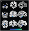
Results: The patients with MDD showed decreased strength in positive dFC between the left CM/SF and brainstem and between the left SF and left thalamus; they showed decreased strength in negative dFC between the left CM and right superior frontal gyrus (p < 0.05, family-wise error-corrected). We found significant positive correlations between age at onset and the mean positive strength of dFC in the left CM/brainstem in patients with MDD.
Limitations: The definitions of amygdalar subregions were based on a cytoarchitectonic delineation, and the temporal resolution of the fMRI was slow (repetition time = 2 s).
Conclusion: These findings confirm the distinct dynamic functional pathway of amygdalar subregions in MDD and suggest that the limbic-cortical-striato-pallido-thalamic circuitry plays a crucial role in the early stages of MDD.
Conflict of interest statement
Figures


References
-
- LeDoux JE. Emotion circuits in the brain. Annu Rev Neurosci. 2000;23:155–84. - PubMed
-
- Pessoa L. On the relationship between emotion and cognition. Nat Rev Neurosci. 2008;9:148–58. - PubMed
-
- van Eijndhoven P, van Wingen G, van Oijen K, et al. Amygdala volume marks the acute state in the early course of depression. Biol Psychiatry. 2009;65:812–8. - PubMed
-
- Frodl T, Meisenzahl EM, Zetzsche T, et al. Larger amygdala volumes in first depressive episode as compared to recurrent major depression and healthy control subjects. Biol Psychiatry. 2003;53:338–44. - PubMed
Publication types
MeSH terms
LinkOut - more resources
Full Text Sources
