Review
. 2018 Nov 14;39(43):3847-3854.
doi: 10.1093/eurheartj/ehy346.

Pulsatile arterial haemodynamics in heart failure

Affiliations
Review

Pulsatile arterial haemodynamics in heart failure

Thomas Weber et al. Eur Heart J. .

Abstract

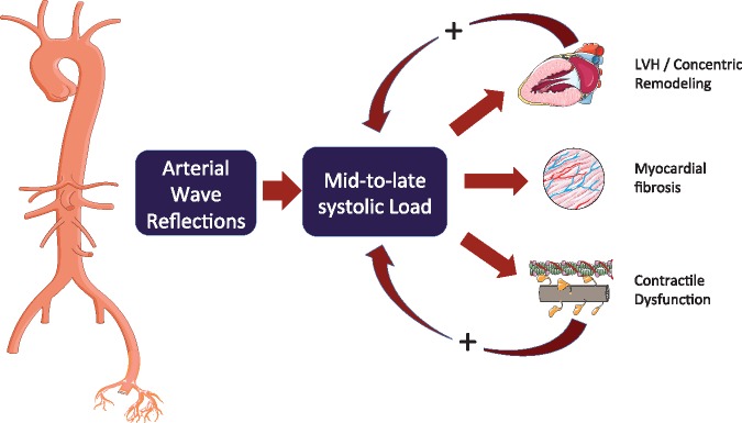
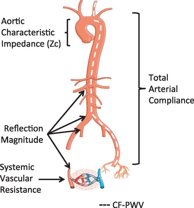
Due to the cyclic function of the human heart, pressure and flow in the circulation are pulsatile rather than continuous. Addressing pulsatile haemodynamics starts with the most convenient measurement, brachial pulse pressure, which is widely available, related to development and treatment of heart failure (HF), but often confounded in patients with established HF. The next level of analysis consists of central (rather than brachial) pressures and, more importantly, of wave reflections. The latter are closely related to left ventricular late systolic afterload, ventricular remodelling, diastolic dysfunction, exercise capacity, and, in the long-term, the risk of new-onset HF. Wave reflection may also represent a suitable therapeutic target. Treatments for HF with preserved and reduced ejection fraction, based on a reduction of wave reflection, are emerging. A full understanding of ventricular-arterial coupling, however, requires dedicated analysis of time-resolved pressure and flow signals, which can be readily accomplished with contemporary non-invasive imaging and modelling techniques. This review provides a summary of our current understanding of pulsatile haemodynamics in HF.


Figures

Figure 1
Figure 1
Assessment of pulsatile haemodynamics—overview. Top line: recording of signal-averaged radial or brachial pressure waveforms with tonometry or brachial cuff. Second line: following calibration with brachial pressures, aortic waveforms are calculated with a transfer function (TF). Pulse waveform analysis, based on pressure signals alone, yields measures of the first (P1) and second (P2) systolic peaks for computation of augmented pressure and augmentation index (AP, AIx). Third line: flow waveforms are obtained, either with Doppler recording of LV outflow (which equals aortic inflow), or as model-derived flow or triangular flow as a proxy. Bottom line: combined and time-aligned analysis of a pressure–flow pair is used for wave separation analysis, wave intensity analysis, and other analytical approaches (Courtesy of Bernhard Hametner, modified from Parragh et al. and from Hametner et al.14). DBP, diastolic blood pressure; ED, ejection duration; MAP, mean arterial pressure; PP, pulse pressure; PWA, pulse waveform analysis, RM, reflection magnitude; SBP, systolic blood pressure; WIA, wave intensity analysis; WSA, wave separation analysis.
Figure 2
Figure 2
Wave separation analysis vs. wave intensity analysis. Identification of forward-travelling and backward travelling waves in the proximal aorta using wave intensity analysis. The method is based on the assessment of changes in pressure (P) and flow velocity (left panels), which can be multiplied to compute instantaneous wave intensity (left bottom panel). The right panel shows forward and backward wave intensity curves, which can be analysed to identify the timing and magnitude of key wave fronts: early systolic forward compression wave (dark blue), late systolic forward suction wave (light blue), and backward compression wave (red). The cumulative sum of forward and backward pressure changes (dP, used to computed wave intensity) are superimposed on the right y-axis. These curves (Pf, Pb) are equivalent to forward and backward waves obtained via classic wave separation analysis. It can be seen that wave intensity tends to markedly under-represent reflected waves, because it emphasizes rapid changes in pressure and flow (high frequencies), whereas reflected waves are enriched in lower frequencies. BCW, backward compression wave; FCW, forward compression wave; FEW, forward expansion suction wave.
Figure 3
Figure 3
Time-resolved myocardial wall stress. The first panel shows the ejection-phase aortic pressure and myocardial wall stress (MWS) profiles. The second panel shows the time-resolved relative myocardial geometry (ratio of wall volume to cavity volume) that correlates with wall stress via the Laplace law; the first, second, and last thirds of systole are shown in blue, dotted red, and black lines, respectively. The third panel shows the ejection-phase myocardial wall stress, and the fourth panel shows pressure–MWS relation. It can be seen that myocardial wall stress peaks in early systole and subsequently decreases, even in the context of increasing pressure. This is due to a mid-systolic shift in the pressure–stress relation (dashed arrow) which favours lower MWS for any given pressure. This shift is due to the geometric reconfiguration of the LV (decreased cavity volume relative to LV wall volume) and is impaired in the presence of reductions in LV ejection fraction, concentric geometric remodelling, and reduced early systolic ejection (reduced early-phase ejection fraction). LV, left ventricular.
Figure 4
Figure 4
Wave reflections increase late systolic left ventricle load, which favours left ventricular remodelling and myocardial dysfunction. However, the effect of wave reflection on myocardial load is modulated by contraction pattern and the time course of myocardial wall stress. Left ventricles in which the mid-systolic shift in the pressure–stress relation is impaired (due to a reduced ejection fraction, concentric geometric remodelling, and/or reduced early systolic ejection) fail to protect cardiomyocytes against the load induced by wave reflections in late systole, a period of vulnerability to load. This may represent a vicious cycle that favours development and furthers progression of heart failure. Modified from Chirinos. LVH, left ventricular hypertrophy.
Figure 5
Figure 5
Anatomic origin of arterial properties that impact left ventricular afterload. Although arterial load results from complex interaction between various arterial segments, in general, specific loading patterns can be attributed to anatomic sites. cf-PWV, a measure of large artery wall stiffness, is also shown, although this is not a measure of LV load per se. Modified from Chirinos and Segers. cf-PWV, carotid-femoral pulse wave velocity.

References

    1. Ponikowski P, Voors AA, Anker SD, Bueno H, Cleland JG, Coats AJ, Falk V, Gonzalez-Juanatey JR, Harjola VP, Jankowska EA, Jessup M, Linde C, Nihoyannopoulos P, Parissis JT, Pieske B, Riley JP, Rosano GM, Ruilope LM, Ruschitzka F, Rutten FH, van der Meer P; ESC Scientific Document Group. 2016 ESC Guidelines for the diagnosis and treatment of acute and chronic heart failure: the Task Force for the diagnosis and treatment of acute and chronic heart failure of the European Society of Cardiology (ESC) Developed with the special contribution of the Heart Failure Association (HFA) of the ESC. Eur Heart J 2016;37:2129–2200. - PubMed
    1. Rapsomaniki E, Timmis A, George J, Pujades-Rodriguez M, Shah AD, Denaxas S, White IR, Caulfield MJ, Deanfield JE, Smeeth L, Williams B, Hingorani A, Hemingway H.. Blood pressure and incidence of twelve cardiovascular diseases: lifetime risks, healthy life-years lost, and age-specific associations in 1.25 million people. Lancet 2014;383:1899–1911. - PMC - PubMed
    1. Williamson JD, Supiano MA, Applegate WB, Berlowitz DR, Campbell RC, Chertow GM, Fine LJ, Haley WE, Hawfield AT, Ix JH, Kitzman DW, Kostis JB, Krousel-Wood MA, Launer LJ, Oparil S, Rodriguez CJ, Roumie CL, Shorr RI, Sink KM, Wadley VG, Whelton PK, Whittle J, Woolard NF, Wright JT Jr, Pajewski NM, Group SR. Intensive vs standard blood pressure control and cardiovascular disease outcomes in adults aged >/=75 years: a randomized clinical trial. J Am Med Assoc 2016;315:2673–2682. - PMC - PubMed
    1. Townsend RR, Wilkinson IB, Schiffrin EL, Avolio AP, Chirinos JA, Cockcroft JR, Heffernan KS, Lakatta EG, McEniery CM, Mitchell GF, Najjar SS, Nichols WW, Urbina EM, Weber T; American Heart Association Council on Hypertension. Recommendations for improving and standardizing vascular research on arterial stiffness: a scientific statement from the American Heart Association. Hypertension 2015;66:698–722. - PMC - PubMed
    1. Parragh S, Hametner B, Bachler M, Kellermair J, Eber B, Wassertheurer S, Weber T.. Determinants and covariates of central pressures and wave reflections in systolic heart failure. Int J Cardiol 2015;190:308–314. - PubMed