. 2018 Aug 21;46(14):7206-7220.
doi: 10.1093/nar/gky541.

Bacillus subtilis RarA modulates replication restart

Affiliations

Bacillus subtilis RarA modulates replication restart

Begoña Carrasco et al. Nucleic Acids Res. .

Abstract

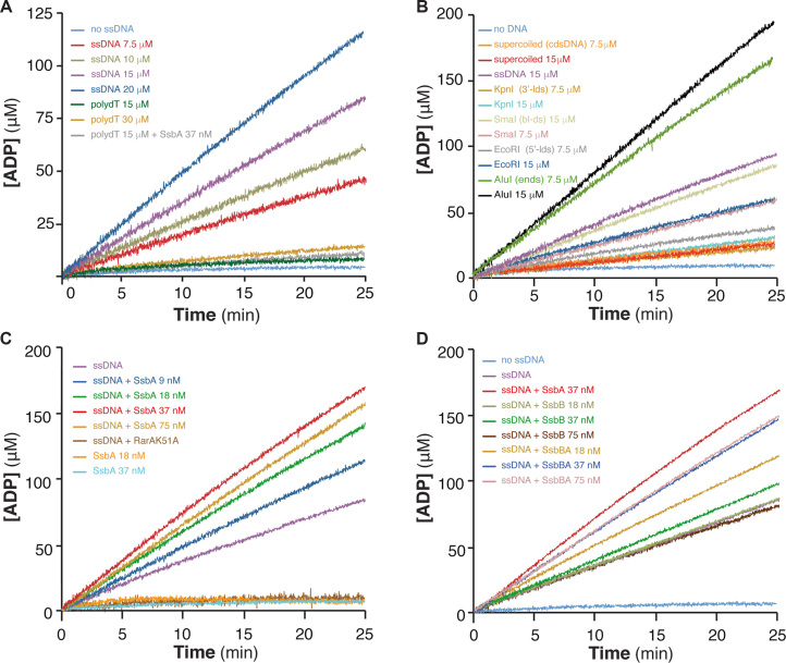
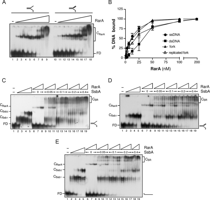
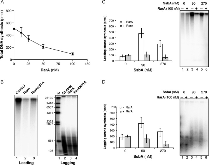
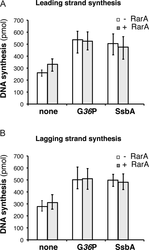
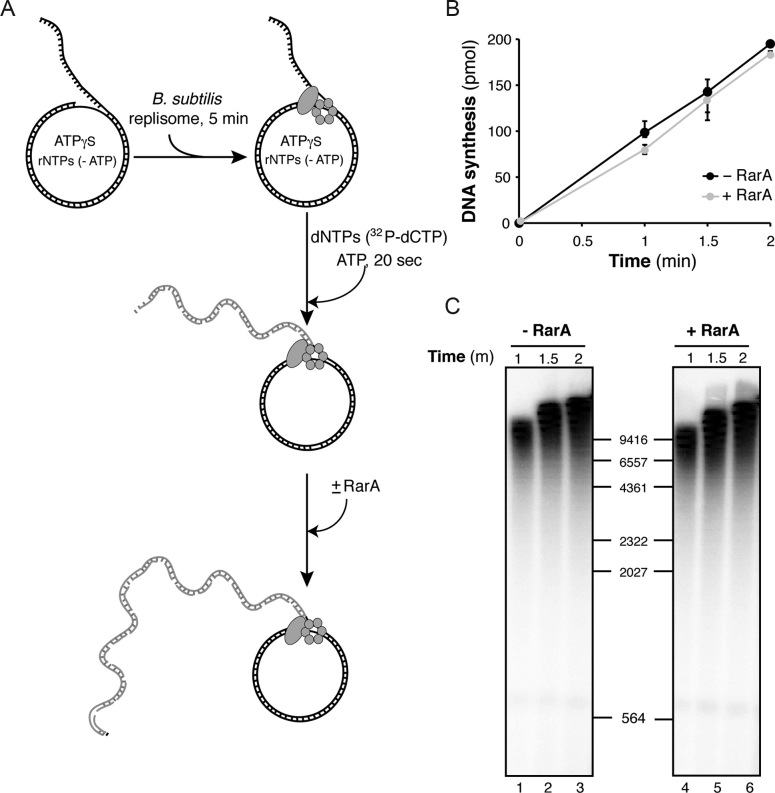
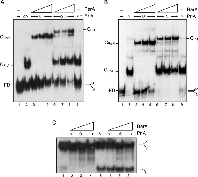
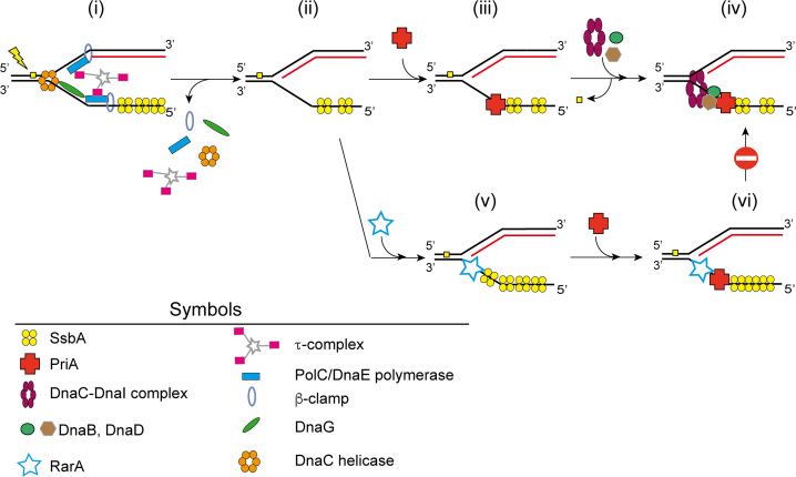
The ubiquitous RarA/Mgs1/WRNIP protein plays a crucial, but poorly understood role in genome maintenance. We show that Bacillus subtilis RarA, in the apo form, preferentially binds single-stranded (ss) over double-stranded (ds) DNA. SsbA bound to ssDNA loads RarA, and for such recruitment the amphipathic C-terminal domain of SsbA is required. RarA is a DNA-dependent ATPase strongly stimulated by ssDNA-dsDNA junctions and SsbA, or by dsDNA ends. RarA, which may interact with PriA, does not stimulate PriA DNA unwinding. In a reconstituted PriA-dependent DNA replication system, RarA inhibited initiation, but not chain elongation. The RarA effect was not observed in the absence of SsbA, or when the host-encoded preprimosome and the DNA helicase are replaced by proteins from the SPP1 phage with similar function. We propose that RarA assembles at blocked forks to maintain genome integrity. Through its interaction with SsbA and with a preprimosomal component, RarA might impede the assembly of the replicative helicase, to prevent that recombination intermediates contribute to pathological DNA replication restart.


Figures

Figure 1.
Figure 1.
RarA ATPase activity is stimulated by dsDNA-ssDNA junctions and dsDNA ends. (A) Stimulation by ssDNA containing secondary structures. RarA (25 nM) was incubated with increasing concentrations of circular 3199-nt ssDNA (7.5 to 20 μM) or linear 80-nt polydT ssDNA (15 and 30 μM) in buffer B containing 5 mM ATP, and the ATPase activity was measured (25 min, 37°C). The ATPase activity of RarA was also measured in the absence of ssDNA (no ssDNA, light blue) and in the presence of polydT and SsbA (gray). (B) Stimulation by dsDNA ends. RarA (25 nM) was incubated with two concentrations (7.5 and 15 μM) of various duplex DNA substrates: supercoiled 3199-bp dsDNA [cdsDNA], or dsDNA linearized with EcoRI [5′-ldsDNA], KpnI [3′-ldsDNA], SmaI [bl-dsDNA] or AluI [dsDNA-ends]. (C) Stimulation by SsbA bound to ssDNA. RarA (25 nM) was incubated with circular 3199-nt ssDNA (15 μM) and increasing concentrations of SsbA (9–75 nM). No ATPase activity is detected in the absence of ssDNA but presence of SsbA (18 nM and 37 nM, orange and blue) or when RarA was replaced by RarAK51A (ssDNA+K51A, dark brown). (D) ATPAse is stimulated by the C-terminal end of SsbA. RarA (25 nM) was incubated with circular 3199-nt ssDNA (15 μM) and increasing SsbB or SsbBA concentrations (18–75 nM). As a control, ATPase activity of RarA in the absence of ssDNA (no ssDNA, blue), in the presence of only ssDNA (magenta), or with ssDNA and SsbA (red) is shown. The amount of ATP hydrolysed was calculated as described (see Materials and Methods). Representative graphics are shown and quantification of the results are expressed as the mean ± SEM of >3 independent experiments (see Supplementary Table S2).
Figure 2.
Figure 2.
Binding affinity of RarA to different DNA substrates. (A) Binding of RarA to forked DNA and to a replicated fork (a replication fork with a fully synthesized leading-strand end and a gap in the lagging strand) determined by EMSA. [γ32P]-radiolabelled DNA (0.4 nM) was incubated with increasing concentrations of RarA (from 1.5 to 200 nM) in buffer B. Protein-DNA complexes were separated as described in methods. (B) RarA binding affinity for [γ32P]-dsDNA, [γ32P]-ssDNA, [γ32P]-fork or a [γ32P]-replicated fork was quantified from EMSA analysis. The results are expressed as the mean ± SEM of >3 independent experiments. (CE) Cooperative binding of RarA and SsbA to forked DNA, replicated fork or ssDNA. The indicated combinations of SsbA (0.05–0.4 nM) and RarA (3–12 nM) were incubated with [γ-32P]-fork (C), [γ-32P]-replicated fork (D) or [γ-32P]-ssDNA (E) in buffer B. Protein–DNA complexes were analysed by native PAGE and autoradiography. Abbreviations: FD, free DNA, CSsb1 and CSsb2, SsbA-DNA complexes; CRarA, RarA–DNA complex and CSR, SsbA–DNA–RarA ternary complexes.
Figure 3.
Figure 3.
SsbA-dependent RarA-mediated inhibition of B. subtilis PriA-dependent DNA replication. (A) Total DNA synthesis obtained in the presence of increasing RarA concentrations (15 min, 37°C). Reaction mixes contained all replisome components (preprimosomal proteins [PriA, DnaB, DnaD, DnaI), DnaC, DnaG, SsbA, τ-complex, β, PolC, DnaE), the indicated RarA concentration, template DNA, rNTPs, dNTPs and [α-32P]-dCTP and [α-32P]-dGTP. An enzyme mix consisting of all proteins except SsbA was generated and added to a substrate mix composed of template DNA, rNTPs, dNTPs, and SsbA. Then, samples were placed at 37°C. (B) Visualization of products obtained in the presence of 100 nM RarA or RarAK51A (15 min, 37°C). In the presence of [α-32P]-dCTP very large DNA fragments derived from rolling circle leading strand DNA synthesis is observed. A parallel reaction in the presence of [α-32P]-dGTP renders visible the small Okazaki fragments due to lagging strand DNA synthesis. Quantification of leading (C) and lagging strand (D) synthesis in the absence/presence of 100nM RarA and the indicated SsbA concentrations (15 min, 37°C). The quantification of the results is expressed as the mean ± SEM of six independent experiments. On the right part, a representative alkaline gel visualized by autoradiography showing the products of the DNA synthesis obtained in the presence or absence of RarA and SsbA.
Figure 4.
Figure 4.
RarA does not inhibit SPP1 DNA replication. Quantification of leading (A) and lagging (B) strand synthesis obtained in standard SPP1 rolling circle DNA replication assays in the absence or in the presence of 100 nM RarA. Reaction mixes contained the SPP1 replisome, which is composed by SPP1 preprimosomal proteins (G38P and G39P) and DNA helicase G40P, and host proteins (DnaG, τ-complex, β, PolC and DnaE). The SPP1 replisome works with both SSB proteins (SsbA or G36P) and the effect of RarA on reactions having either viral G36P or host SbsA was tested. An enzyme mix consisting of all proteins except the SSB was generated, and added to a substrate mix composed of template DNA, rNTPs, dNTPs, and the indicated SSB (none, 30 nM G36P, or, 90 nM SsbA). Then reactions were placed at 37°C and incubated for 10 min. Leading strand synthesis was quantified by [α-32P]-dCTP incorporation and lagging strand synthesis by [α-32P]-dGTP incorporation. The results are expressed as the mean ± SEM of >3 independent experiments.
Figure 5.
Figure 5.
RarA has no effect on ongoing DNA replication. (A) Scheme of the experimental design. The B. subtilis replisome was assembled on the DNA in the absence of RarA and in the presence of limiting ATPγS and then DNA replication was started by dNTP (including [α-32P]-dCTP) and ATP addition. After 20 s of initiating the reaction, 100 nM RarA was added or not, and reactions were continued for the indicated times. (B) Quantification of leading strand synthesis (mean ± SEM of >3 independent experiments). (C) The leading strand DNA products obtained in one of these assays are visualized by denaturing gel electrophoresis and autoradiography.
Figure 6.
Figure 6.
RarA may interact with PriA but does not stimulate its helicase activity. (A and B) Simultaneous binding of RarA and PriA to a replicated fork. PriA (2.5 nM [A], and 5 nM [B]) and RarA (25–100 nM) were incubated with [γ-32P]-replicated fork (0.4 nM in molecules) in buffer C. Protein-DNA complexes were analysed by native PAGE and autoradiography. Abbreviations: FD, free DNA, CPriA, PriA-DNA complex; CRarA, RarA–DNA complex and CPR, PriA–DNA–RarA ternary complex. (C) RarA does not stimulate the helicase activity of PriA. The indicated combinations of PriA (5 nM) and RarA (25, 50, 100 nM) were incubated with the helicase substrate ([γ-32P]-fork) in buffer E (30 min, 30°C). Products were separated after deproteinization by PAGE and visualized by autoradiography. RarA was unable to unwind this substrate under these experimental conditions (lanes 2–4).
Figure 7.
Figure 7.
Model of RarA action on blocked forks. (i) When a lesion (filled yellow square) blocks DNA replication, DNA synthesis is stopped and the replisome might disassemble. (ii) SsbA binds to the lagging strand of a stalled replication fork. (iii) By protein interaction PriA is recruited. (iv) PriA bound to the lagging strand recruits, by protein-protein interaction, DnaB and DnaD and the DnaC-DnaI helicase–loader complex to the stalled fork. Then the damage is repaired. The helicase is then activated by DnaI release and subsequent preprimosome disassembly. DnaC recruits DnaG primase and the replisome and DNA replication can restart (not depicted). (v and vi) If the DNA damage is not removed, SsbA loads RarA at the stalled fork. The loading of PriA is not avoided, but the SsbA-RarA-PriA-DNA complex impedes the recruitment of the replisome and initiation of DNA synthesis is inhibited.

Similar articles

Cited by

References

    1. Yoshimura A., Seki M., Enomoto T.. The role of WRNIP1 in genome maintenance. Cell Cycle. 2017; 16:515–521. - PMC - PubMed
    1. Barre F.X., Soballe B., Michel B., Aroyo M., Robertson M., Sherratt D.. Circles: the replication-recombination-chromosome segregation connection. Proc. Natl. Acad. Sci. U.S.A. 2001; 98:8189–8195. - PMC - PubMed
    1. Lau I.F., Filipe S.R., Soballe B., Okstad O.A., Barre F.X., Sherratt D.J.. Spatial and temporal organization of replicating Escherichia coli chromosomes. Mol. Microbiol. 2003; 49:731–743. - PubMed
    1. Shibata T., Hishida T., Kubota Y., Han Y.W., Iwasaki H., Shinagawa H.. Functional overlap between RecA and MgsA (RarA) in the rescue of stalled replication forks in Escherichia coli. Genes Cells. 2005; 10:181–191. - PubMed
    1. Lestini R., Michel B.. UvrD controls the access of recombination proteins to blocked replication forks. EMBO J. 2007; 26:3804–3814. - PMC - PubMed

Publication types

MeSH terms

LinkOut - more resources