Review
. 2018 Sep;75(17):3181-3191.
doi: 10.1007/s00018-018-2855-3. Epub 2018 Jun 15.

Coordination of microtubule acetylation and the actin cytoskeleton by formins

Affiliations
Review

Coordination of microtubule acetylation and the actin cytoskeleton by formins

Jaime Fernández-Barrera et al. Cell Mol Life Sci. 2018 Sep.

Abstract

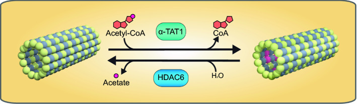
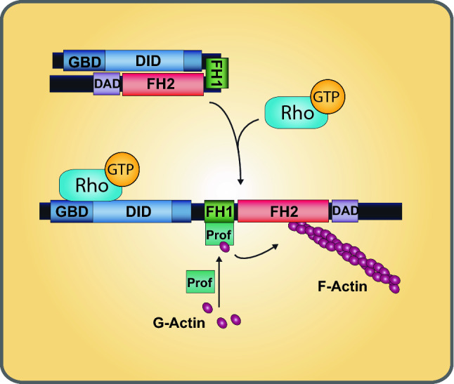
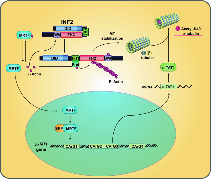
The acetylation of the lysine 40 residue of α-tubulin was described more than 30 years ago and has been the subject of intense research ever since. Although the exact function of this covalent modification of tubulin in the cell remains unknown, it has been established that tubulin acetylation confers resilience to mechanical stress on the microtubules. Formins have a dual role in the fate of the actin and tubulin cytoskeletons. On the one hand, they catalyze the formation of actin filaments, and on the other, they bind microtubules, act on their stability, and regulate their acetylation and alignment with actin fibers. Recent evidence indicates that formins coordinate the actin cytoskeleton and tubulin acetylation by modulating the levels of free globular actin (G-actin). G-actin, in turn, controls the activity of the myocardin-related transcription factor-serum response factor transcriptional complex that regulates the expression of the α-tubulin acetyltransferase 1 (α-TAT1) gene, which encodes the main enzyme responsible for tubulin acetylation. The effect of formins on tubulin acetylation is the combined result of their ability to activate α-TAT1 gene transcription and of their capacity to regulate microtubule stabilization. The contribution of these two mechanisms in different formins is discussed, particularly with respect to INF2, a formin that is mutated in hereditary human renal and neurodegenerative disorders.

Keywords: Actin homeostasis; Formins; INF2; Microtubules; Myocardin-related transcription factor; Serum response factor; Tubulin acetylation; α-Tubulin acetyltransferase 1.


Figures

Fig. 1
Fig. 1
Enzymes involved in tubulin acetylation and deacetylation. Tubulin acetylation refers to the transfer of the acetyl group from acetyl-coenzyme A to the K40 residue of α-tubulin. This modification is catalyzed by α-TAT1 in mammals, whereas its reverse reaction is catalyzed by the deacetylase HDAC6. Note that the K40 residue of α-tubulin is located within the microtubule lumen
Fig. 2
Fig. 2
Regulation of diaphanous-related formins and domain organization of distinct formins. The autoinhibitory effect of the DID–DAD interaction in the diaphanous-related formin group is released through binding of a specific Rho-family GTPase in its active GTP-loaded form. In the open conformation of formins, the FH1 domain recruits profilin (Prof), which, in turn, brings actin monomers close to the FH2 domain for actin polymerization
Fig. 3
Fig. 3
Domain organization of the indicated formins. The domain involved in MT stabilization is indicated. The molecules are not drawn to scale
Fig. 4
Fig. 4
Affinity constants of MRTF and different formins for G-actin. Compilation of the dissociation constants of MRTF and of the DAD/WH2 of INF2, mDia1 and FMNL3 for G-actin, and dissociation constants for the DID–DAD interaction in these formins
Fig. 5
Fig. 5
Schematic of the proposed model of INF2 function on microtubule acetylation. Given that the affinity of the WH2/DAD (W/D) of INF2 for G-actin is much higher than for the DID, INF2 may sense the increase of the levels of G-actin better than other formins. Since, in the case of INF2, the binding of G-actin to the DAD/WH2 releases INF2 from autoinhibition, increased levels of free G-actin result in INF2-mediated actin polymerization and, consequently, a decrease in free G-actin. This decrease allows MRTF to enter the nucleus and associate with SRF to direct the transcription of the α-TAT1 gene and other target genes. The increased levels of α-TAT1 mRNA produce more α-TAT1 and, subsequently, MT acetylation on the K40 residue of α-tubulin augments. In addition, INF2 contributes to tubulin acetylation via MT stabilization by forming part of a large protein complex that stabilizes MTs in an active Rho GTPAse-dependent manner

References

    1. Borisy G, Heald R, Howard J, Janke C, Musacchio A, Nogales E. Microtubules: 50 years on from the discovery of tubulin. Nat Rev Cell Mol Biol. 2016;17:322–328. doi: 10.1038/nrm.2016.45. - DOI - PMC - PubMed
    1. Muroyama A, Lechler T. Microtubule organization, dynamics and functions in differentiated cells. Development. 2017;144:3012–3021. doi: 10.1242/dev.153171. - DOI - PMC - PubMed
    1. Etienne-Manneville S. Microtubules in cell migration. Annu Rev Cell Dev Biol. 2013;29:471–499. doi: 10.1146/annurev-cellbio-101011-155711. - DOI - PubMed
    1. Bartolini F, Gundersen GG. Generation of noncentrosomal microtubule arrays. J Cell Sci. 2006;119:4155. doi: 10.1242/jcs.03227. - DOI - PubMed
    1. Hammond JW, Cai D, Verhey KJ. Tubulin modifications and their cellular functions. Curr Opin Cell Biol. 2008;20:71–76. doi: 10.1016/j.ceb.2007.11.010. - DOI - PMC - PubMed

Publication types

LinkOut - more resources