

Real-time, intraoperative detection of residual breast cancer in lumpectomy cavity walls using a novel cathepsin-activated fluorescent imaging system
- PMID: 29948401
- PMCID: PMC6193492
- DOI: 10.1007/s10549-018-4845-4
Real-time, intraoperative detection of residual breast cancer in lumpectomy cavity walls using a novel cathepsin-activated fluorescent imaging system
Abstract
Purpose: Obtaining tumor-free surgical margins is critical to prevent recurrence in breast-conserving surgery but it remains challenging. We assessed the LUM Imaging System for real-time, intraoperative detection of residual tumor.
Methods: Lumpectomy cavity walls and excised specimens of breast cancer lumpectomy patients were assessed with the LUM Imaging System (Lumicell, Inc., Wellesley MA) with and without intravenous LUM015, a cathepsin-activatable fluorescent agent. Fluorescence at potential sites of residual tumor was evaluated with a sterile hand-held probe, displayed on a monitor and correlated with histopathology.
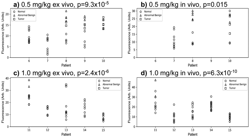
Results: Background autofluorescence was assessed in excised specimens from 9 patients who did not receive LUM015. In vivo lumpectomy cavities and excised specimens were then imaged in 15 women undergoing breast cancer surgery who received no LUM015, 0.5, or 1 mg/kg LUM015 (5 women per dose). Among these, 11 patients had invasive carcinoma with ductal carcinoma in situ (DCIS) and 4 had only DCIS. Image acquisition took 1 s for each 2.6-cm-diameter surface. No significant background normal breast fluorescence was identified. Elevated fluorescent signal was seen from invasive cancers and DCIS. Mean tumor-to-normal signal ratios were 4.70 ± 1.23 at 0.5 mg/kg and 4.22 ± 0.9 at 1.0 mg/kg (p = 0.54). Tumor was distinguished from normal tissue in pre-and postmenopausal women and readings were not affected by breast density. Some benign tissues produced fluorescent signal with LUM015.
Conclusion: The LUM Imaging System allows rapid identification of residual tumor in the lumpectomy cavity of breast cancer patients and may reduce rates of positive margins.
Keywords: Breast cancer; Image-guided surgery; Intraoperative tumor detection; Lumpectomy surgery.
Conflict of interest statement
Authors D. Strasfeld and J. Ferrer have received remuneration and stock ownership from Lumicell, Inc. All other authors declare that he/she have no conflicts of interest.
Figures
References
-
- Veronesi U, Cascinelli N, Mariani L, et al. (October 17, 2002) Twenty-year follow-up of a randomized study comparing breast-conserving surgery with radical mastectomy for early breast cancer. N Engl J Med 347(16):1227–32. - PubMed
-
- Clarke M, Collins R, Darby S, et al. (2005) Early Breast Cancer Trialists' Collaborative Group (EBCTCG). Effects of radiotherapy and of differences in the extent of surgery for early breast cancer on local recurrence and 15-year survival: an overview of the randomised trials. Lancet 366:2087–106. - PubMed
MeSH terms
Substances
Grants and funding
LinkOut - more resources
Full Text Sources
Other Literature Sources
Medical
Miscellaneous
