Review
. 2018 Sep;131(5):727-734.
doi: 10.1007/s10265-018-1050-9. Epub 2018 Jun 12.

The cellular machineries responsible for the division of endosymbiotic organelles

Affiliations
Review

The cellular machineries responsible for the division of endosymbiotic organelles

Yamato Yoshida. J Plant Res. 2018 Sep.

Erratum in

Abstract

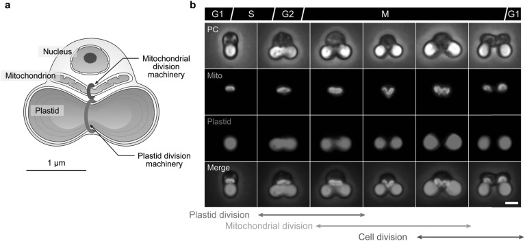
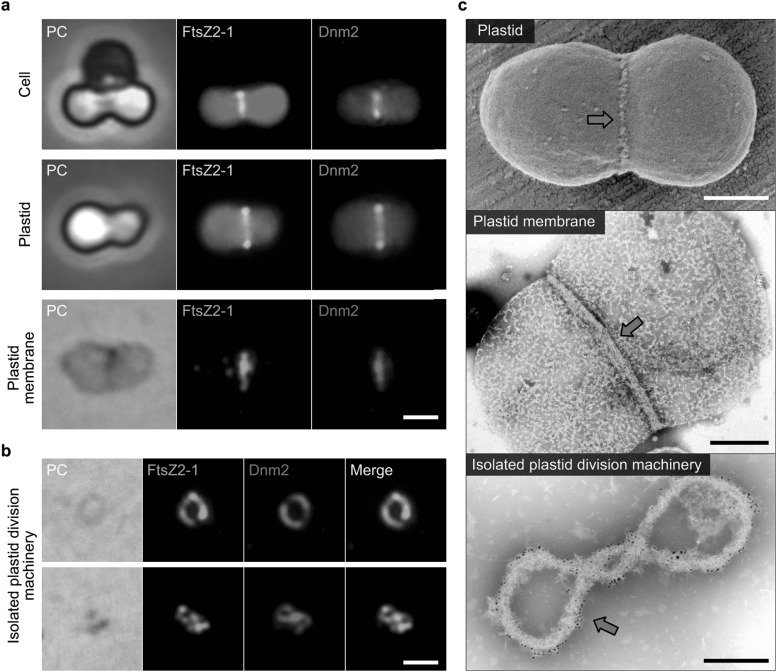
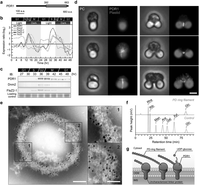
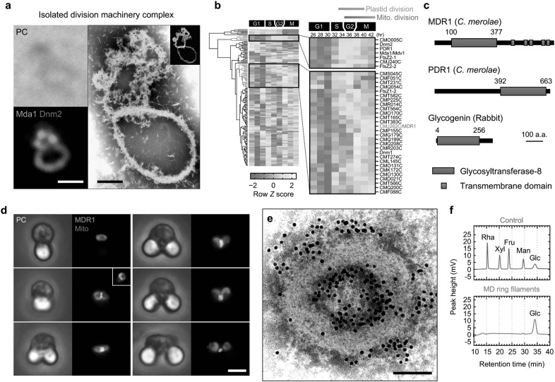
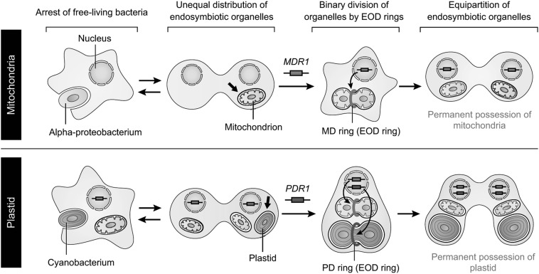
Chloroplasts (plastids) and mitochondria evolved from endosymbiotic bacteria. These organelles perform vital functions in photosynthetic eukaryotes, such as harvesting and converting energy for use in biological processes. Consistent with their evolutionary origins, plastids and mitochondria proliferate by the binary fission of pre-existing organelles. Here, I review the structures and functions of the supramolecular machineries driving plastid and mitochondrial division, which were discovered and first studied in the primitive red alga Cyanidioschyzon merolae. In the past decade, intact division machineries have been isolated from plastids and mitochondria and examined to investigate their underlying structure and molecular mechanisms. A series of studies has elucidated how these division machineries assemble and transform during the fission of these organelles, and which of the component proteins generate the motive force for their contraction. Plastid- and mitochondrial-division machineries have important similarities in their structures and mechanisms despite sharing no component proteins, implying that these division machineries evolved in parallel. The establishment of these division machineries might have enabled the host eukaryotic ancestor to permanently retain these endosymbiotic organelles by regulating their binary fission and the equal distribution of resources to daughter cells. These findings provide key insights into the establishment of endosymbiotic organelles and have opened new avenues of research into their evolution and mechanisms of proliferation.

Keywords: Chloroplast division; Endosymbiotic organelle; MDR1; Mitochondrial division; PDR1.


Figures

Fig. 1
Fig. 1
The primitive unicellular red alga C. merolae. a Schematic representation of a dividing C. merolae cell. b Sequential process of plastid, mitochondrial, and cell division in C. merolae. Mitochondria were imaged following immunostaining with an anti-mitochondrial-porin antibody, and the plastids were imaged by chlorophyll autofluorescence. PC, phase contrast. Scale bar, 1 µm. Images in b were reproduced and modified, with permission, from Yoshida et al. (2013)
Fig. 2
Fig. 2
Isolation of plastid division machineries from dividing plastids in C. merolae cells. a Phase-contrast and immunofluorescence images of the FtsZ (FtsZ2-1: yellow/green) and dynamin (Dnm2: orange) rings of whole cells (top), isolated plastids (middle), and plastid membranes (bottom). Scale bar, 1 µm. b Isolated plastid division machineries. Scale bar, 1 µm. c Electron micrographs of a dividing plastid, a plastid membrane, and isolated plastid division machinery. Red arrows indicate plastid division machineries. Scale bars, 500 nm (top and middle) and 200 nm (bottom). Images were reproduced and modified, with permission, from Yoshida et al. (2006) and Yoshida et al. (2010)
Fig. 3
Fig. 3
Identification of PLASTID-DIVIDING RING1 (PDR1) in C. merolae. a Domain architecture of CMR358C/PDR1. b Expression of PDR1, Dnm2, FtsZ2-1, and FtsZ2-2 throughout the cell cycle, determined using a time-course transcriptome dataset from synchronized C. merolae cells (Fujiwara et al. 2009). c Protein levels of PDR1, Dnm2, and FtsZ2-1 throughout the cell cycle. d Immunofluorescence images of PDR1 in C. merolae cells. Scale bar, 1 µm. e Immuno-EM of PDR1 in the plastid division machinery. Many more PDR1 proteins were detected in the less-condensed regions of PD ring filaments than in the solid region (insets), suggesting that PDR1 protein molecules are associated with the whole PD ring filament, not just the surface. Scale bars, 100 nm (left) and 50 nm (right). f Fluorometric detection of the acid-hydrolysis products of the PD ring filament fraction using HPLC. g A schematic representation of PD ring filament biosynthesis by PDR1. Images were reproduced and modified, with permission, from Yoshida et al. (2010) and Yoshida (2018)
Fig. 4
Fig. 4
Identification of MITOCHONDRION-DIVIDING RING1 (MDR1) in C. merolae. a Isolated division machinery complexes containing the mitochondrial division machinery (green) and the plastid division machinery (red). Scale bars, 500 nm (left) and 200 nm (right). b Hierarchical clustering analysis of genes characterized using a proteomic analysis of isolated division machinery complexes. c Domain architecture of MDR1, PDR1, and glycogenin. d Phase-contrast and immunofluorescence images of MDR1. Scale bar, 1 µm. e Immuno-EM of isolated overdeveloped mitochondrial division machinery. Overdeveloped mitochondrial division machineries were isolated from mitochondrial-division-arrested C. merolae cells, which were obtained by treatment with a DNA synthesis inhibitor (see Yoshida et al. for more detail). In this image, 71% of the immunogold signals indicating the presence of MDR1 are located on the inner periphery region of the isolated mitochondrial division machinery. Scale bar, 100 nm. f Component analysis of the purified MD ring filaments using HPLC. Images were reproduced and modified, with permission, from Yoshida et al. (2017)
Fig. 5
Fig. 5
Proposed model for the evolutionary processes underlying the establishment of endosymbiotic organelles. See text and Yoshida et al. (2017) for more details. Schematic representations were reproduced and modified, with permission, from Yoshida et al. (2017)

Similar articles

Cited by

References

    1. Bleazard W, McCaffery JM, King EJ, et al. The dynamin-related GTPase Dnm1 regulates mitochondrial fission in yeast. Nat Cell Biol. 1999;1:298–304. doi: 10.1038/13014. - DOI - PMC - PubMed
    1. Chen C, MacCready JS, Ducat DC, Osteryoung KW. The molecular machinery of chloroplast division. Plant Physiol. 2018;176:138–151. doi: 10.1104/pp.17.01272. - DOI - PMC - PubMed
    1. Friedman JR, Nunnari J. Mitochondrial form and function. Nature. 2014;505:335–343. doi: 10.1038/nature12985. - DOI - PMC - PubMed
    1. Fujiwara T, Misumi O, Tashiro K, et al. Periodic gene expression patterns during the highly synchronized cell nucleus and organelle division cycles in the unicellular red alga Cyanidioschyzon merolae. DNA Res. 2009;16:59–72. doi: 10.1093/dnares/dsn032. - DOI - PMC - PubMed
    1. Gao H, Kadirjan-Kalbach D, Froehlich JE, Osteryoung KW. ARC5, a cytosolic dynamin-like protein from plants, is part of the chloroplast division machinery. Proc Natl Acad Sci U S A. 2003;100:4328–4333. doi: 10.1073/pnas.0530206100. - DOI - PMC - PubMed

LinkOut - more resources