. 2018 Aug;38(6):1293-1303.
doi: 10.1007/s10571-018-0597-2. Epub 2018 Jun 13.

Upregulation of miR-199a-5p Protects Spinal Cord Against Ischemia/Reperfusion-Induced Injury via Downregulation of ECE1 in Rat

Affiliations

Upregulation of miR-199a-5p Protects Spinal Cord Against Ischemia/Reperfusion-Induced Injury via Downregulation of ECE1 in Rat

Ning Bao et al. Cell Mol Neurobiol. 2018 Aug.

Abstract

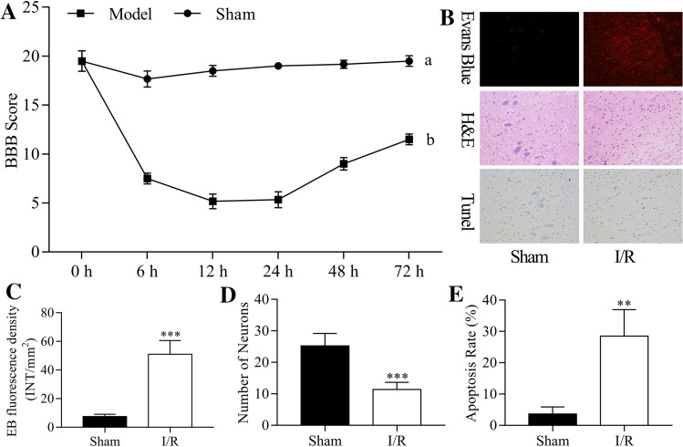
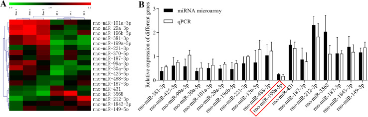
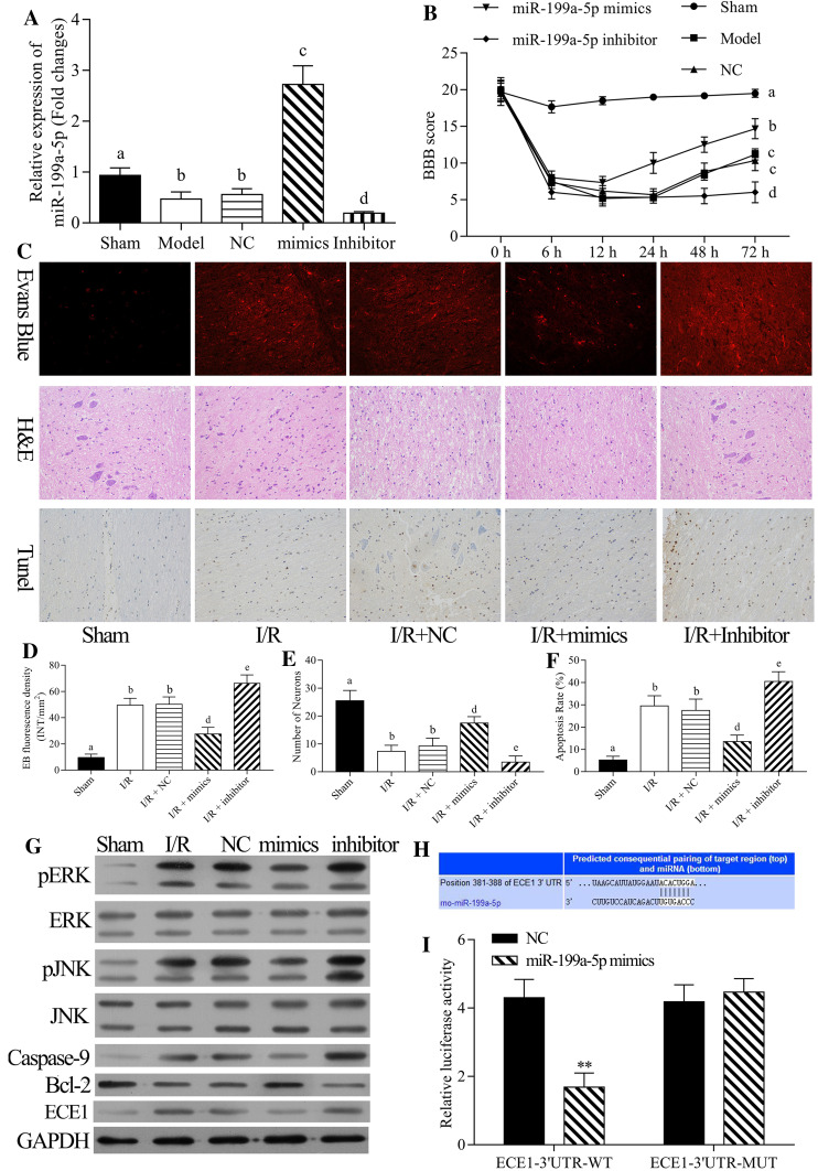
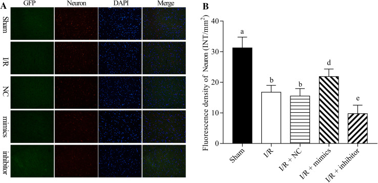
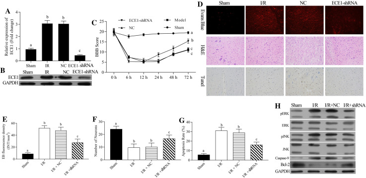
Ischemia-reperfusion (I/R)-induced spinal cord injury can cause apoptotic damage and subsequently act as a blood-spinal cord barrier damage. MicroRNAs (miRNAs) contributed to the process of I/R injury by regulating their target mRNAs. miR-199a-5p is involved in brain and heart I/R injury; however, its function in the spinal cord is not yet completely clarified. In this study, we investigated the role of miR-199a-5p on spinal cord I/R via the endothelin-converting enzyme 1, especially the apoptosis pathway. In the current study, the rat spinal cord I/R injury model was established, and the Basso Beattie Bresnahan scoring, Evans blue staining, HE staining, and TUNEL assay were used to assess the I/R-induced spinal cord injury. The differentially expressed miRNAs were screened using microarray. miR-199a-5p was selected by unsupervised hierarchical clustering analysis. The dual-luciferase reporter assay was used for detecting the regulatory effects of miR-199a-5p on ECE1. In addition, neuron expression was detected by immunostaining assay, while the expressions of p-ERK, ERK, p-JNK, JNK, caspase-9, Bcl-2, and ECE1 were evaluated by Western blot. The results indicated the successful establishment of the I/R-induced spinal cord injury model; the I/R induced the damage to the lower limb motor. Furthermore, 18 differentially expressed miRNAs were detected in the I/R group compared to the sham group, and miR-199a-5p protected the rat spinal cord injury after I/R. Moreover, miR-199a-5p negatively regulated ECE1, and silencing the ECE1 gene also protected the rat spinal cord injury after I/R. miR-199a-5p or silencing of ECE1 also regulated the expressions of caspase-9, Bcl-2, p-JNK, p-ERK, and ECE1 in rat spinal cord injury after I/R. Therefore, we demonstrated that miR-199a-5p might protect the spinal cord against I/R-induced injury by negatively regulating the ECE1, which could aid in developing new therapeutic strategies for I/R-induced spinal cord injury.

Keywords: Apoptosis; Blood–spinal cord barrier; ECE1; Ischemia/reperfusion; MiR-199a-5p.


Conflict of interest statement

Ning Bao, Bo Fang, Huangwei Lv, Yanhua Jiang, Fengshou Chen, Zhilin Wang, and Hong Ma declare that they have no financial and personal relationships with people or organizations that can inappropriately influence the current study; there is no professional or other personal interest of any nature or kind in any product, service and/or company that could be construed as influencing the position presented in, or the review of the manuscript entitled “Upregulation of miR-199a-5p protects spinal cord against ischemia/reperfusion-induced injury via downregulation of ECE1 in rat.”

Figures

Fig. 1
Fig. 1
I/R-induced damage of lower limb motor. The rat spinal cord I/R injury model was established. a The lower limb motor function was assessed using BBB scoring criteria at 0, 6, 12, 24, 48, and 72 h post-injury. b EB fluorescence, number of neurons, and cell apoptosis were detected by EB, H&E, and TUNEL assays at 24 h post-injury, respectively. c EB fluorescence density was quantitatively analyzed by EB assay. d The number of neurons was counted according to H&E staining. e The rate of apoptosis was calculated according to the TUNEL assay
Fig. 2
Fig. 2
MiRNAs expression in the rat spinal cord after I/R. The rat spinal cord I/R injury model was established. a Heat map of the differentially expressed miRNAs in the sham (n = 6) and I/R groups (n = 6). Each column indicates the expression profile of a sample; and red is high expression, green is low expression. b miR-199a-5p expression was analyzed by qRT-PCR assay in the sham and model groups
Fig. 3
Fig. 3
Effects of miR-199a-5p on the rat spinal cord injury after I/R. The rat spinal cord I/R injury model was established and transfected with negative control (NC), miR-199a-5p mimic, and miR-199a-5p inhibitor, respectively. a The mRNA expression level of miR-199a-5p was detected by qRT-PCR assay. b The lower limb motor function of rats was analyzed by BBB scoring criteria at spinal cord I/R injury at 0, 6, 12, 24, 48, and 72 h post-reperfusion. c EB fluorescence, number of neurons, and cell apoptosis were measured by EB, H&E, and TUNEL assays at 24 h post-injury, respectively. d EB fluorescence density, e number of neurons, and f cell apoptosis were analyzed quantitatively. g The protein expression levels of p-ERK, ERK, p-JNK, JNK, caspase-9, Bcl-2, and ECE1 were measured by Western blot assay. h The putative complementary site of miR-199a-5p and ECE1 was obtained based on TargetScan prediction. Cells were transfected with wild-type ECE1 and mutant-type ECE1, and NC or miR-199a-5p mimic, respectively. The luciferase intensity was measured by dual-luciferase reporter assay. **P < 0.01 vs. NC group. ac P < 0.05 vs. sham, I/R and NC, respectively
Fig. 4
Fig. 4
miR-199a-5p inducing neuron proliferation in I/R-induced spinal cord injury rats. The rat spinal cord I/R injury model was established and transfected with negative control (NC), miR-199a-5p mimic, and miR-199a-5p inhibitor, respectively. a The immunostaining assay was performed to assess the expression of neuron-specific beta III tubulin. b Neuron-specific beta III tubulin expression was quantified. DAPI indicates the nucleus. ac P < 0.05 vs. sham, I/R and NC, respectively
Fig. 5
Fig. 5
Silencing of ECE1 exerting a protective effect on the rat spinal cord injury after I/R. The rat spinal cord I/R injury model was established and transfected with negative control (NC) and shRNA-ECE1s, respectively. (a) The mRNA expression level of ECE1 was accessed by qRT-PCR assay. b The protein expression level of ECE1 was analyzed by Western blot assay. c The lower limb motor function of rats was evaluated by BBB scoring criteria of spinal cord I/R injury at 0, 6, 12, 24, 48, and 72 h post-reperfusions. d EB fluorescence, number of neurons, and cell apoptosis were measured by EB, H&E, and TUNEL assays at 24 h post-injury, respectively. e EB fluorescence density, f number of neurons, and g cell apoptosis were analyzed quantitatively, respectively. h Expressions of p-ERK, ERK, p-JNK, JNK, caspase-9, Bcl-2, and ECE1 were measured by Western blot assay. ac P < 0.05 vs. sham, I/R and NC, respectively

References

    1. Anttila V, Haapanen H, Yannopoulos F, Herajarvi J, Anttila T, Juvonen T (2016) Review of remote ischemic preconditioning: from laboratory studies to clinical trials. Scand Cardiovasc J 50(5–6):355–361. 10.1080/14017431.2016.1233351 - DOI - PubMed
    1. Anwar MA, Al Shehabi TS, Eid AH (2016) Inflammogenesis of secondary spinal cord injury. Front Cell Neurosci 10:98. 10.3389/fncel.2016.00098 - DOI - PMC - PubMed
    1. Arsava EM, Gurer G, Gursoy-Ozdemir Y, Karatas H, Dalkara T (2009) A new model of transient focal cerebral ischemia for inducing selective neuronal necrosis. Brain Res Bull 78(4–5):226–231. 10.1016/j.brainresbull.2008.11.005 - DOI - PubMed
    1. Azzarone M, De Troia A, Iazzolino L, Nabulsi B, Tecchio T (2016) Hybrid treatment of acute abdominal aortic thrombosis presenting with paraplegia. Ann Vasc Surg 33:228.e225-228. 10.1016/j.avsg.2015.10.041 - DOI - PubMed
    1. Basso DM, Beattie MS, Bresnahan JC (1995) A sensitive and reliable locomotor rating scale for open field testing in rats. J Neurotrauma 12(1):1–21. 10.1089/neu.1995.12.1 - DOI - PubMed

LinkOut - more resources