

The Hypothalamic-Pituitary-Adrenal Axis and Serotonin Metabolism in Individual Brain Nuclei of Mice with Genetic Disruption of the NK1 Receptor Exposed to Acute Stress
- PMID: 29948553
- PMCID: PMC11481836
- DOI: 10.1007/s10571-018-0594-5
The Hypothalamic-Pituitary-Adrenal Axis and Serotonin Metabolism in Individual Brain Nuclei of Mice with Genetic Disruption of the NK1 Receptor Exposed to Acute Stress
Abstract
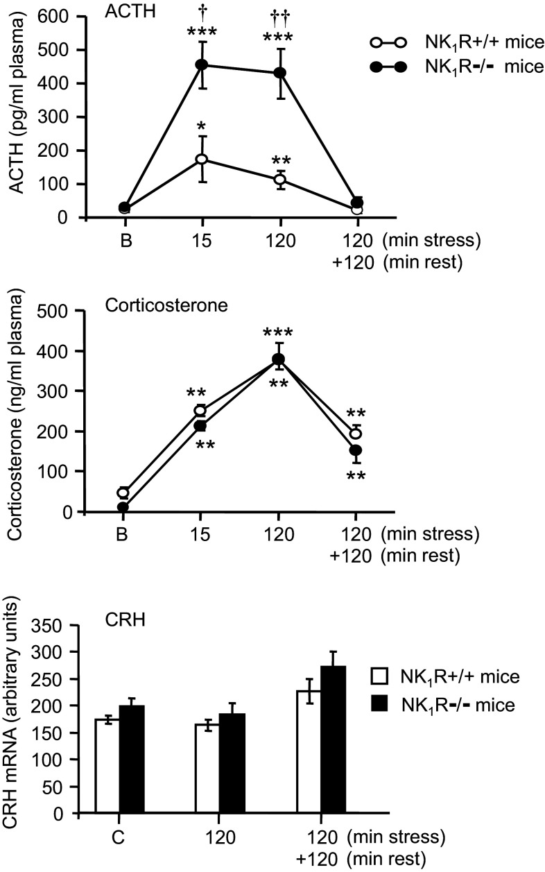
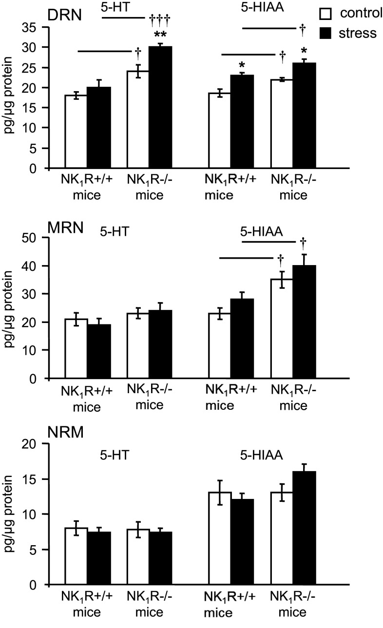
Mice lacking the substance P (SP) neurokinin-1 (NK1) receptor (NK1R-/-mice) were used to investigate whether SP affects serotonin (5-HT) function in the brain and to assess the effects of acute immobilisation stress on the hypothalamic-pituitary-adrenocortical (HPA) axis and 5-HT turnover in individual brain nuclei. Basal HPA activity and the expression of hypothalamic corticotropin-releasing hormone (CRH) in wild-type (WT)- and NK1R-/- mice were identical. Stress-induced increases in plasma ACTH concentration were considerably higher in NK1R-/- mice than in WT mice while corticosterone concentrations were equally elevated in both mouse lines. Acute stress did not alter the expression of CRH. In the dorsal raphe nucleus (DRN), basal 5-HT turnover was increased in NK1R-/- mice and a 15 min stress further magnified 5-HT utilisation in this region. In the frontoparietal cortex, medial prefrontal cortex, central nucleus of amygdala, and the hippocampal CA1 region, stress increased 5-HT and/or 5-hydroxyindoleacetic acid (5-HIAA) concentrations to a similar extent in WT and NK1R-/- mice. 5-HT turnover in the hypothalamic paraventricular nucleus was not affected by stress, but stress induced similar increases in 5-HT and 5-HIAA in the ventromedial and dorsomedial hypothalamic nuclei in WT and NK1R-/- mice. Our findings indicate that NK1 receptor activation suppresses ACTH release during acute stress but does not exert sustained inhibition of the HPA axis. Genetic deletion of the NK1 receptor accelerates 5-HT turnover in DRN under basal and stress conditions. No differences between the responses of serotonergic system to acute stress in WT and NK1R-/- mice occur in forebrain nuclei linked to the regulation of anxiety and neuroendocrine stress responses.
Keywords: ACTH; Brain; CRH; Mouse; NK1 receptor; Serotonin; Stress; Substance P.
Conflict of interest statement
The authors have no conflict of interest to declare.
Figures
Similar articles
-
Serotonin activates the hypothalamic-pituitary-adrenal axis via serotonin 2C receptor stimulation.J Neurosci. 2007 Jun 27;27(26):6956-64. doi: 10.1523/JNEUROSCI.2584-06.2007. J Neurosci. 2007. PMID: 17596444 Free PMC article.
-
Substance P is involved in terminating the hypothalamo- pituitary-adrenal axis response to acute stress through centrally located neurokinin-1 receptors.Stress. 2000 May;3(3):209-20. doi: 10.3109/10253890009001125. Stress. 2000. PMID: 10938582
-
Acute glucocorticoid pretreatment suppresses stress-induced hypothalamic-pituitary-adrenal axis hormone secretion and expression of corticotropin-releasing hormone hnRNA but does not affect c-fos mRNA or fos protein expression in the paraventricular nucleus of the hypothalamus.J Neuroendocrinol. 2003 Nov;15(11):1075-83. doi: 10.1046/j.1365-2826.2003.01100.x. J Neuroendocrinol. 2003. PMID: 14622438
-
Studies on the neuroendocrine role of serotonin.Dan Med Bull. 2007 Nov;54(4):266-88. Dan Med Bull. 2007. PMID: 18208678 Review.
-
HPA axis responsiveness to stress: implications for healthy aging.Exp Gerontol. 2011 Feb-Mar;46(2-3):90-5. doi: 10.1016/j.exger.2010.08.023. Epub 2010 Sep 9. Exp Gerontol. 2011. PMID: 20833240 Free PMC article. Review.
Cited by
-
Resilience to the effects of social stress on vulnerability to developing drug addiction.World J Psychiatry. 2022 Jan 19;12(1):24-58. doi: 10.5498/wjp.v12.i1.24. eCollection 2022 Jan 19. World J Psychiatry. 2022. PMID: 35111578 Free PMC article. Review.
References
-
- Arborelius L, Owens MJ, Plotsky PM, Nemeroff CB (1999) The role of corticotropin-releasing factor in depression and anxiety disorders. J Endocrinol 160:1–12 - PubMed
-
- Bernardis LL, Bellinger LL (1998) The dorsomedial hypothalamic nucleus revisited: 1998 update. Proc Soc Exp Biol Med 218:284–306 - PubMed
MeSH terms
Substances
LinkOut - more resources
Full Text Sources
Other Literature Sources
Miscellaneous
