Comparative Study
. 2019 Apr;21(2):257-268.
doi: 10.1007/s11307-018-1212-0.

SNAPshots of the MCHR1: a Comparison Between the PET-Tracers [18F]FE@SNAP and [11C]SNAP-7941

Affiliations
Comparative Study

SNAPshots of the MCHR1: a Comparison Between the PET-Tracers [18F]FE@SNAP and [11C]SNAP-7941

Cécile Philippe et al. Mol Imaging Biol. 2019 Apr.

Abstract

Purpose: The melanin-concentrating hormone receptor 1 (MCHR1) has become an important pharmacological target, since it may be involved in various diseases, such as diabetes, insulin resistance, and obesity. Hence, a suitable positron emission tomography radiotracer for the in vivo assessment of the MCHR1 pharmacology is imperative. The current paper contrasts the extensive in vitro, in vivo, and ex vivo assessments of the radiotracers [18F]FE@SNAP and [11C]SNAP-7941 and provides comprehensive information about their biological and physicochemical properties. Furthermore, it examines their suitability for first-in-man imaging studies.

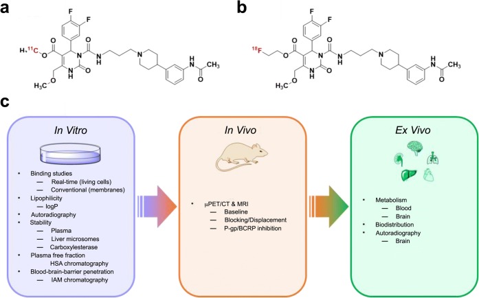
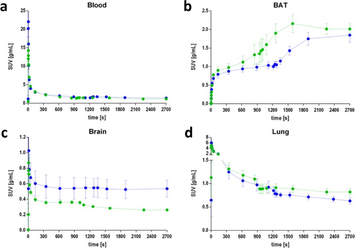
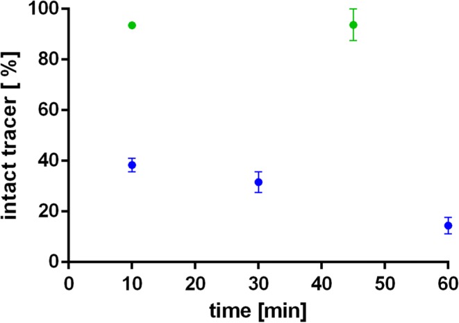
Procedures: Kinetic real-time cell-binding studies with [18F]FE@SNAP and [11C]SNAP-7941 were conducted on adherent Chines hamster ovary (CHO-K1) cells stably expressing the human MCHR1 and MCHR2. Small animal imaging studies on mice and rats were performed under displacement and baseline conditions, as well as after pretreatment with the P-glycoprotein/breast cancer resistant protein inhibitor tariquidar. After the imaging studies, detailed analyses of the ex vivo biodistribution were performed. Ex vivo metabolism was determined in rat blood and brain and analyzed at various time points using a quantitative radio-HPLC assay.

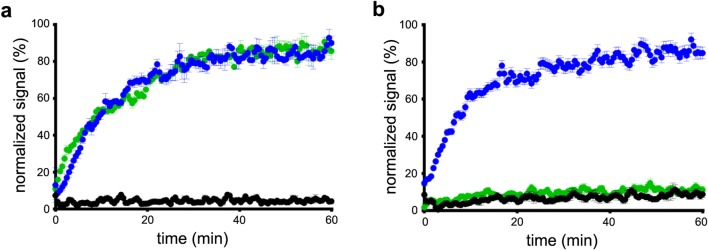
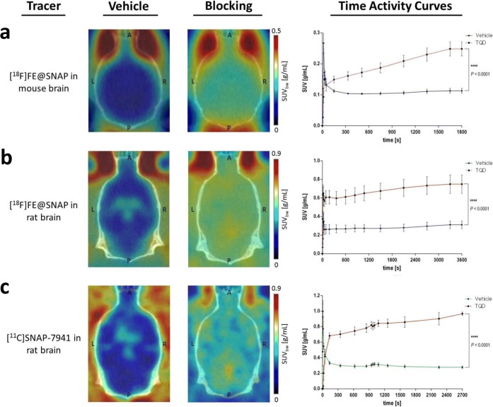
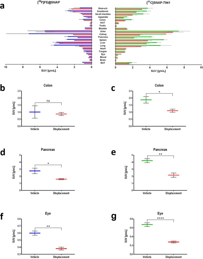
Results: [11C]SNAP-7941 demonstrates high uptake on CHO-K1-hMCHR1 cells, whereas no uptake was detected for the CHO-K1-hMCHR2 cells. In contrast, [18F]FE@SNAP evinced binding to CHO-K1-hMCHR1 and CHO-K1-hMCHR2 cells. Imaging studies with [18F]FE@SNAP and [11C]SNAP-7941 showed an increased brain uptake after tariquidar pretreatment in mice, as well as in rats, and exhibited a significant difference between the time-activity curves of the baseline and blocking groups. Biodistribution of both tracers demonstrated a decreased uptake after displacement. [11C]SNAP-7941 revealed a high metabolic stability in rats, whereas [18F]FE@SNAP was rapidly metabolized.

Conclusions: Both radiotracers demonstrate appropriate imaging properties for the MCHR1. However, the pronounced metabolic stability as well as superior selectivity and affinity of [11C]SNAP-7941 underlines the decisive superiority over [18F]FE@SNAP.

Keywords: Ex vivo; In vitro; In vivo; MCHR1; PET; Small animal imaging; [11C]SNAP-7941; [18F]FE@SNAP.


Conflict of interest statement

The authors declare that there is no conflict of interest.

Figures

Fig. 1
Fig. 1
Chemical structures of a [11C]SNAP-7941 and b [18F]FE@SNAP andc corresponding in vitro, in vivo, and ex vivo experiments. IAM: immobilized artificial membrane; HAS: human serum albumin; P-gp: P-glycoprotein; BCRP: breast cancer resistance protein.
Fig. 2
Fig. 2
Depiction of the association kinetics of [11C]SNAP-7941 (green) and [18F]FE@SNAP (blue) on adherent a CHO-K1-hMCHR1 and b CHO-K1-hMCHR2 cells. The black curve indicates the accumulation of both tracers to native CHO-K1 cells. Data are displayed as mean ± SEM from independent experiments (n ≥ 3, each performed as a quadruplicate, 0.05–1000 nM).
Fig. 3
Fig. 3
Representation of axial planes of mouse (a) and rat (b, c) brains under vehicle and blocking conditions with the P-gp/BCRP inhibitor TQD for [18F]FE@SNAP (a, b) and [11C]SNAP-7941 (c). Respective TACs are presented on the right-hand side of each illustration for the dedicated radiotracers [18F]FE@SNAP (blue), [11C]SNAP-7941 (green) under vehicle conditions, and in combination with 15-mg/kg TQD (red). Data are displayed as mean ± SEM from independent experiments (n ≥ 3). Differences among groups were tested using a repeated measures ANOVA (**** = P < 0.0001). If not visible, error bars are within the margin of the symbols.
Fig. 4
Fig. 4
TACs for [18F]FE@SNAP (blue) and [11C]SNAP-7941 (green) before and after displacement with 15 mg/kg (±)-SNAP-7941 in a blood, b BAT, c brain, and d lung, after 900 or 1200 s, respectively. Data are displayed as mean ± SEM from independent experiments (n ≥ 3). If not visible, error bars are within the margin of the symbols.
Fig. 5
Fig. 5
a Representative ex vivo rat biodistribution of [11C]SNAP-7941 and [18F]FE@SNAP and bg selected target regions of both radiotracers [18F]FE@SNAP (blue) and [11C]SNAP-7941 (green) under vehicle conditions and after displacement with 15 mg/kg (±)-SNAP-7941 (red). Data are displayed as mean ± SEM from independent experiments (n ≥ 3). Differences among groups were tested using a two-tailed parametric paired t test (ns = P > 0.05; * = P < 0.05; ** = P < 0.01; **** = P < 0.0001). If not visible, error bars are within the margin of the symbols.
Fig. 6
Fig. 6
Ex vivo metabolic stability of [18F]FE@SNAP (blue) and [11C]SNAP-7941 (green) in rat whole blood over time. Data are displayed as mean ± SEM from independent experiments (n ≥ 3). If not visible, error bars are within the margin of the symbols.

Similar articles

Cited by

References

    1. Bittencourt JC. Anatomical organization of the melanin-concentrating hormone peptide family in the mammalian brain. Gen Comp Endocrinol. 2011;172:185–197. doi: 10.1016/j.ygcen.2011.03.028. - DOI - PubMed
    1. Tadayyon M, Welters HJ, Haynes AC, Cluderay JE, Hervieu G. Expression of melanin-concentrating hormone receptors in insulin-producing cells: MCH stimulates insulin release in RINm5F and CRI-G1 cell-lines. Biochem Biophys Res Commun. 2000;275:709–712. doi: 10.1006/bbrc.2000.3357. - DOI - PubMed
    1. Kokkotou E, Moss AC, Torres D, Karagiannides I, Cheifetz A, Liu S, O'Brien M, Maratos-Flier E, Pothoulakis C. Melanin-concentrating hormone as a mediator of intestinal inflammation. Proc Natl Acad Sci U S A. 2008;105:10613–10618. doi: 10.1073/pnas.0804536105. - DOI - PMC - PubMed
    1. Bradley RL, Kokkotou EG, Maratos-Flier E, Cheatham B. Melanin-concentrating hormone regulates leptin synthesis and secretion in rat adipocytes. Diabetes. 2000;49:1073–1077. doi: 10.2337/diabetes.49.7.1073. - DOI - PubMed
    1. Bradley RL, Mansfield JPR, Maratos-Flier E, Cheatham B. Melanin-concentrating hormone activates signaling pathways in 3T3-L1 adipocytes. Am J Physiol Endocrinol Metab. 2002;283:E584–E592. doi: 10.1152/ajpendo.00161.2002. - DOI - PubMed

Publication types

LinkOut - more resources