. 2018 Sep;137(3):503-513.
doi: 10.1007/s11120-018-0534-8. Epub 2018 Jun 13.

Development of fluorescence quenching in Chlamydomonas reinhardtii upon prolonged illumination at 77 K

Affiliations

Development of fluorescence quenching in Chlamydomonas reinhardtii upon prolonged illumination at 77 K

Lucyna M Wlodarczyk et al. Photosynth Res. 2018 Sep.

Abstract

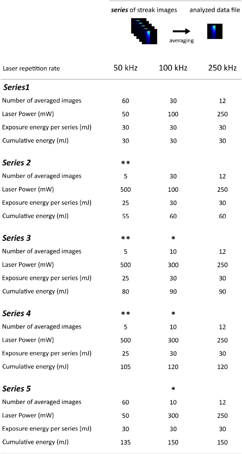
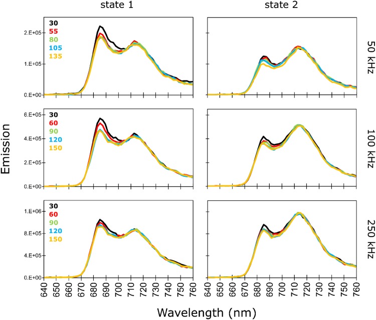
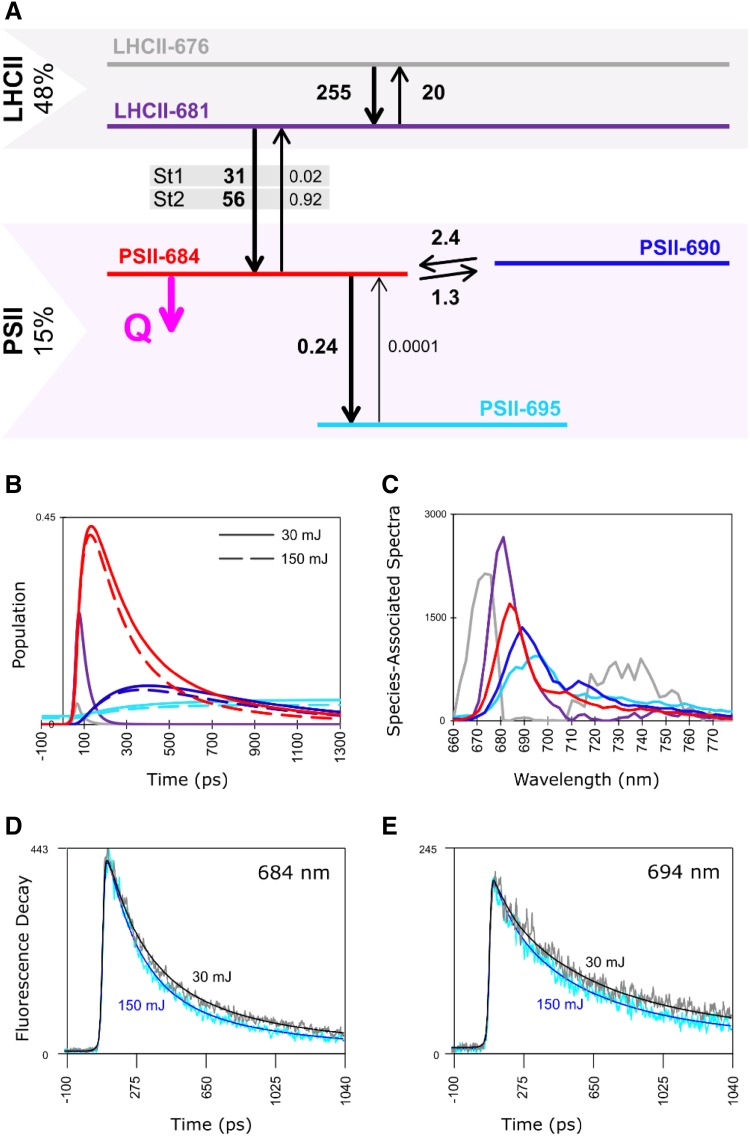
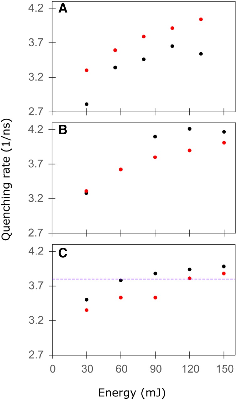
Low-temperature fluorescence measurements are frequently used in photosynthesis research to assess photosynthetic processes. Upon illumination of photosystem II (PSII) frozen to 77 K, fluorescence quenching is observed. In this work, we studied the light-induced quenching in intact cells of Chlamydomonas reinhardtii at 77 K using time-resolved fluorescence spectroscopy with a streak camera setup. In agreement with previous studies, global analysis of the data shows that prolonged illumination of the sample affects the nanosecond decay component of the PSII emission. Using target analysis, we resolved the quenching on the PSII-684 compartment which describes bulk chlorophyll molecules of the PSII core antenna. Further, we quantified the quenching rate constant and observed that as the illumination proceeds the accumulation of the quencher leads to a speed up of the fluorescence decay of the PSII-684 compartment as the decay rate constant increases from about 3 to 4 ns- 1. The quenching on PSII-684 leads to indirect quenching of the compartments PSII-690 and PSII-695 which represent the red chlorophyll of the PSII core. These results explain past and current observations of light-induced quenching in 77 K steady-state and time-resolved fluorescence spectra.

Keywords: Photosystem II; State transitions; Target analysis; Time-resolved fluorescence.


Figures

Fig. 1
Fig. 1
Scheme of data preparation for global and target analysis. Detailed description in the Materials and methods
Fig. 2
Fig. 2
77 K steady-state fluorescence spectra reconstructed from globally analysed time-resolved fluorescence measured upon excitation at 400 nm in C. reinhardtii WT after incubation for 45 min under St1 conditions (left panel) or under St2 conditions (right panel). The spectra were normalized using a scaling factor obtained upon global analysis (more details in Materials and Methods). For each laser repetition rate, state 1 and state 2 spectra have the same vertical axis, and different colours of the spectra represent different cumulative exposure energies indicated in mJ (Fig. 1)
Fig. 3
Fig. 3
Decay-associated spectra (DAS) resulting from global analysis of 77 K time-resolved fluorescence measured upon excitation at 400 nm with laser repetition rate of 100 kHz in C. reinhardtii WT cells after incubation under St1 conditions. Different colours of the spectra represent different cumulative exposure energies indicated in mJ (Fig. 1)
Fig. 4
Fig. 4
Target analysis of 77 K time-resolved fluorescence measured in C. reinhardtii WT cells in St1. The cells were continuously illuminated with 400 nm laser light at 100 kHz repetition rate. a Compartmental model for the LHCII–PSII complex. The population directly after excitation per subunit PSII (15%) and LHCII (48%) is indicated (Table S2). The colour key of the compartments in a is used in b and c. Numbers next to the black arrows indicate rate constants in ns− 1. The natural decay rate constant is 0.2 ns− 1 for all compartments (omitted for clarity). The light-induced quenching rate constant Q (indicated in magenta) increases from 3.3 to 4.2 ns− 1 with cumulative exposure energy 30 to 150 mJ. b Population dynamics in the LHCII–PSII complex with cumulative exposure energy 30 mJ (solid) or 150 mJ (dashed). c Estimated SAS of each compartment. Emission of PSII-690 and PSII-695 above 700 nm is discussed in the Supplementary Information. d Time-resolved emission at 684 nm and e at 694 nm. Traces are scaled to their maximum. Key: accumulated energy 30 mJ (data—grey; fit—black, Q = 3.3 ns− 1), 150 mJ (data—cyan; fit—blue, Q = 4.2 ns− 1)
Fig. 5
Fig. 5
Quenching rate constant Q as a function of the cumulative exposure energy upon prolonged illumination of C. reinhardtii WT cells in St1 (black) or in St2 (red). The laser repetition rate was 50 kHz (a), 100 kHz (b) or 250 kHz (c). Values of Q and error estimates are listed in Table S1. The quenching rate constant of 3.8 ns− 1 estimated in our previous study (Snellenburg et al. 2017) is indicated with a violet dashed line in C

Similar articles

Cited by

References

    1. Allen JF. State transitions—a question of balance. Science. 2003;299:1530–1532. doi: 10.1126/science.1082833. - DOI - PubMed
    1. Allen JF, Bennett J, Steinback KE, Arntzen CJ. Chloroplast protein phosphorylation couples plastoquinone redox state to distribution of excitation energy between photosystems. Nature. 1981;291:25–29. doi: 10.1038/291025a0. - DOI
    1. Allorent G, Tokutsu R, Roach T, et al. A dual strategy to cope with high light in Chlamydomonas reinhardtii. Plant Cell. 2013;25:545–557. doi: 10.1105/tpc.112.108274. - DOI - PMC - PubMed
    1. Andrizhiyevskaya EG, Chojnicka A, Bautista JA, et al. Origin of the F685 and F695 fluorescence in Photosystem II. Photosynth Res. 2005;84:173–180. doi: 10.1007/s11120-005-0478-7. - DOI - PubMed
    1. Bellafiore S, Barneche F, Peltier G, Rochaix J. State transitions and adaptation to changing light require chloroplast thylakoid protein kinase STN7. Nature. 2005;433:2003–2006. doi: 10.1038/nature03286. - DOI - PubMed

MeSH terms

LinkOut - more resources