

Optimization of reconstruction and quantification of motion-corrected coronary PET-CT
- PMID: 29948889
- PMCID: PMC6289874
- DOI: 10.1007/s12350-018-1317-5
Optimization of reconstruction and quantification of motion-corrected coronary PET-CT
Abstract
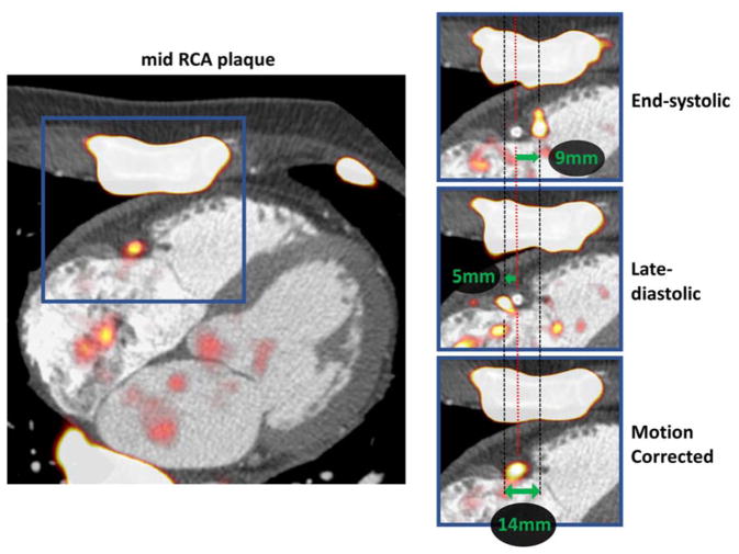
Background: Coronary PET shows promise in the detection of high-risk atherosclerosis, but there remains a need to optimize imaging and reconstruction techniques. We investigated the impact of reconstruction parameters and cardiac motion-correction in 18F Sodium Fluoride (18F-NaF) PET.
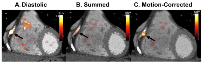
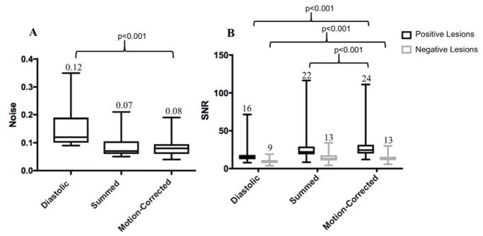
Methods: Twenty-two patients underwent 18F-NaF PET within 22 days of an acute coronary syndrome. Optimal reconstruction parameters were determined in a subgroup of six patients. Motion-correction was performed on ECG-gated data of all patients with optimal reconstruction. Tracer uptake was quantified in culprit and reference lesions by computing signal-to-noise ratio (SNR) in diastolic, summed, and motion-corrected images.
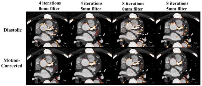
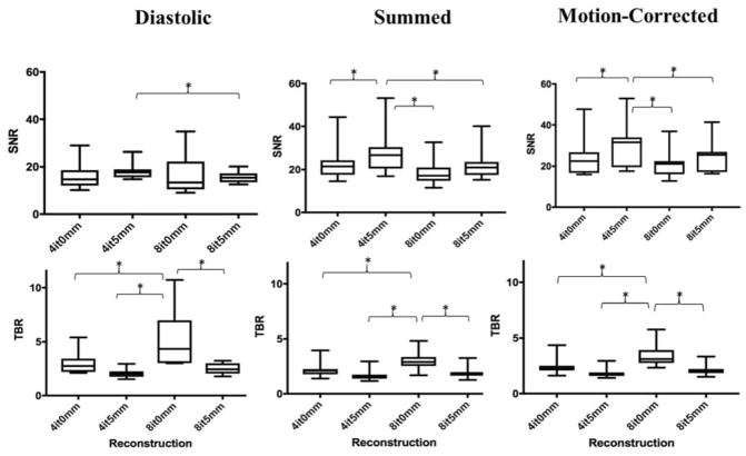
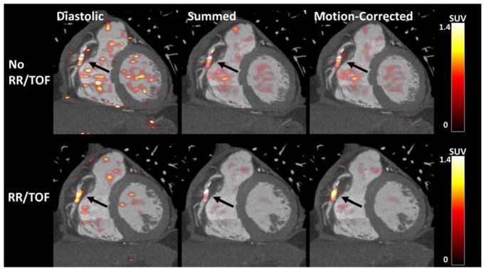
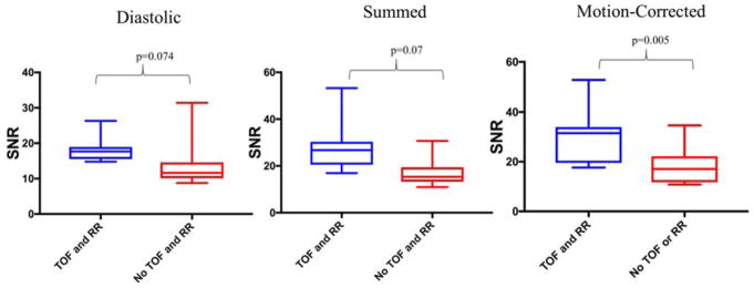
Results: Reconstruction using 24 subsets, 4 iterations, point-spread-function modelling, time of flight, and 5-mm post-filtering provided the highest median SNR (31.5) compared to 4 iterations 0-mm (22.5), 8 iterations 0-mm (21.1), and 8 iterations 5-mm (25.6; all P < .05). Motion-correction improved SNR of culprit lesions (n = 33) (24.5[19.9-31.5]) compared to diastolic (15.7[12.4-18.1]; P < .001) and summed data (22.1[18.9-29.2]; P < .001). Motion-correction increased the SNR difference between culprit and reference lesions (10.9[6.3-12.6]) compared to diastolic (6.2[3.6-10.3]; P = .001) and summed data (7.1 [4.8-11.6]; P = .001).
Conclusions: The number of iterations and extent of post-filtering has marked effects on coronary 18F-NaF PET quantification. Cardiac motion-correction improves discrimination between culprit and reference lesions.
Keywords: Atherosclerosis; Cardiac motion; Computed tomography; Positron emission tomography.
Conflict of interest statement
This research was supported in part by grant 1R01HL135557 from the National Heart, Lung, and Blood Institute/National Institute of Health (NHLBI/NIH). The content is solely the responsibility of the authors and does not necessarily represent the official views of the National Institutes of Health. The study was also supported by a grant (“Cardiac Imaging Research Initiative”) from the Adelson Medical Research Foundation.
David Newby (CH/09/002) and Marc Dweck (FS/14/78) are supported by the British Heart Foundation. David Newby is also the recipient of a Wellcome Trust Senior Investigator Award (WT103782AIA). No other potential conflict of interest relevant to this article was reported.
Figures
Comment in
-
Advances in coronary molecular imaging: Leveraging the power of image processing.J Nucl Cardiol. 2020 Apr;27(2):505-507. doi: 10.1007/s12350-018-1454-x. Epub 2018 Oct 26. J Nucl Cardiol. 2020. PMID: 30367381 Free PMC article.
References
-
- Joshi NJ, Vesey AT, Williams MC, et al. 18F-fluoride positron emission tomography for identification of ruptured and high-risk coronary atherosclerotic plaques: a prospective clinical trial. The Lancet. 2014;383(9918):705–713. - PubMed
-
- Lee JM, Bang J-I, Koo BK, et al. Clinical Relevance of 18F-Sodium Fluoride Positron-Emission Tomography in Noninvasive Identification of High-Risk Plaque in Patients With Coronary Artery Disease. Circ Cardiovasc Imaging. 2017;10(11):e006704. - PubMed
-
- Kitagawa T, Yamamoto H, Toshimitsu S, et al. 18F-sodium fluoride positron emission tomography for molecular imaging of coronary atherosclerosis based on computed tomography analysis. Atherosclerosis. 2017;263:385–392. - PubMed
-
- Rubeaux M, Joshi N, Dweck MR, et al. Motion correction of 18F-sodium fluoride PET for imaging coronary atherosclerotic plaques. J Nucl Med. 2016;57(1):54–59. - PubMed
Publication types
MeSH terms
Substances
Grants and funding
LinkOut - more resources
Full Text Sources
Other Literature Sources
Medical
