. 2018 Jun 27;13(6):e0199683.
doi: 10.1371/journal.pone.0199683. eCollection 2018.

Development of a high-throughput assay to detect antibody inhibition of low pH induced conformational changes of influenza virus hemagglutinin

Affiliations

Development of a high-throughput assay to detect antibody inhibition of low pH induced conformational changes of influenza virus hemagglutinin

Jessica F Trost et al. PLoS One. .

Abstract

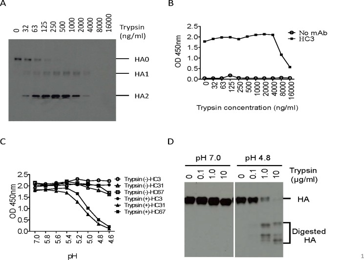
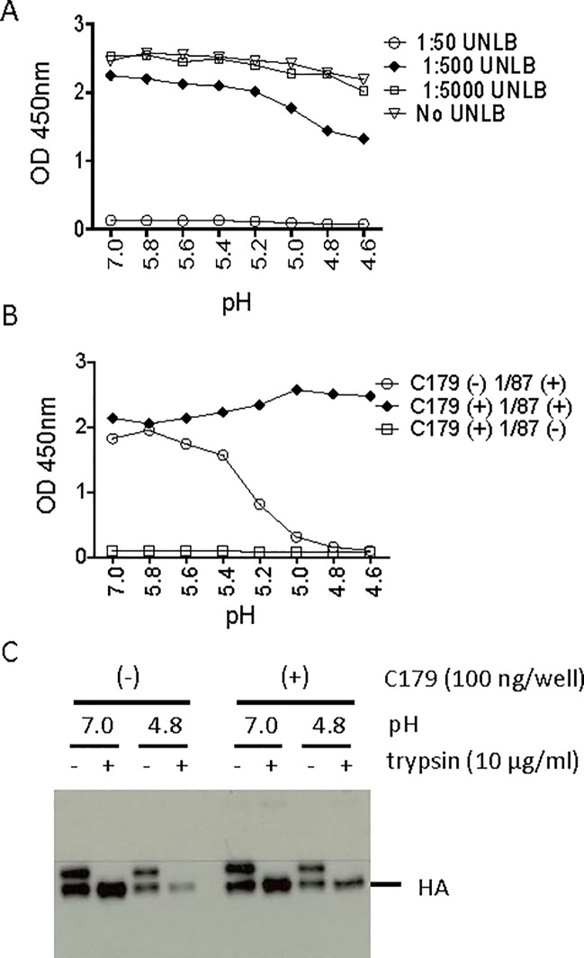
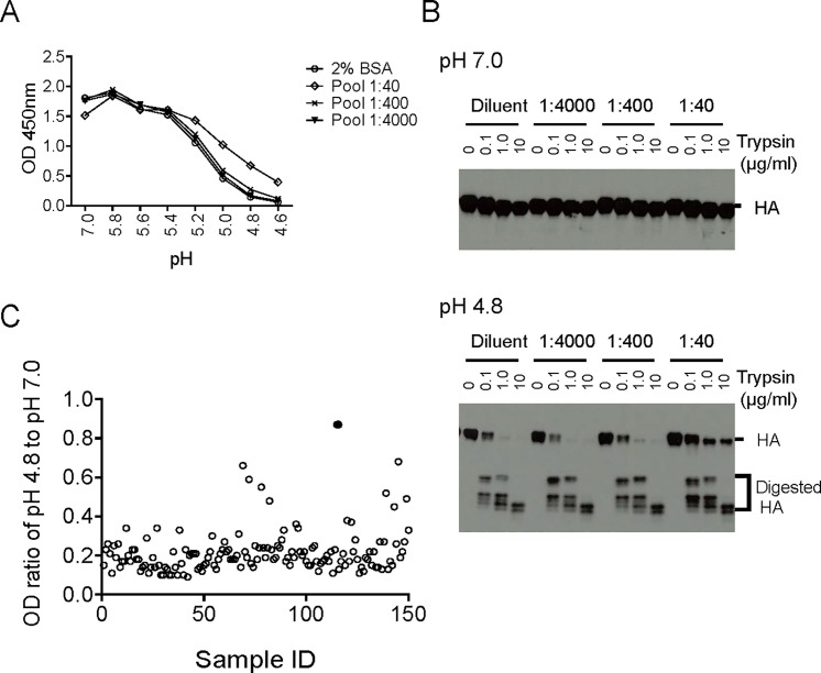
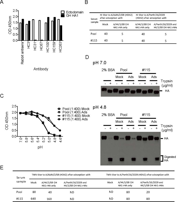
Many broadly neutralizing antibodies (bnAbs) bind to conserved areas of the hemagglutinin (HA) stalk region and can inhibit the low pH induced HA conformational changes necessary for viral membrane fusion activity. We developed and evaluated a high-throughput virus-free and cell-free ELISA based low pH induced HA Conformational Change Inhibition Antibody Detection Assay (HCCIA) and a complementary proteinase susceptibility assay. Human serum samples (n = 150) were tested by HCCIA using H3 recombinant HA. Optical density (OD) ratios of mAb HC31 at pH 4.8 to pH 7.0 ranged from 0.87 to 0.09. Our results demonstrated that low pH induced HA conformational change inhibition antibodies (CCI) neutralized multiple H3 strains after removal of head-binding antibodies. The results suggest that HCCIA can be utilized to detect and characterize CCI in sera, that are potentially broadly neutralizing, and serves as a useful tool for evaluating universal vaccine candidates targeting the HA stalk.


Conflict of interest statement

The authors have declared that no competing interests exist.

Figures

Fig 1
Fig 1. Principle of HCCIA.
A. The linear schematic of the rHA as described [41] with some modifications, not to scale. N-terminal baculovirus GP67 signal peptide, HA ectodomain, mutated thrombin cleavage site from LVPRGS to LVPAGS, foldon, and 6 histidine-Tag. B. The structure shown in the schematic was modified from a published HA structure (PDB No. 1HTM and 4WE4) [–43]. Though trimeric rHA was used in the HCCIA, the H3 rHA is shown as a monomer in Fig 1. In the absence of CCI, H3 rHA would undergo conformational changes at the pH of fusion and HC31 (HC67 for H3 rHA, or 1/87 mAb for H2 rHA) would lose the ability to bind to the rHA, while HC3 (shown in black) would bind to the H3 rHA when in its fusogenic conformation. C. In the presence of CCI (shown in green), H3 rHA would remain “locked” in its metastable pre-fusion conformation and maintain recognition by both HC3 (shown in black) and HC31 (shown in red), as well as HC67 or 1/87 mAb for H2 rHA.
Fig 2
Fig 2. Determination of low pH induced conformational change of H3 rHA on 96-well nickel-coated plate.
Optimization of trypsin concentration for cleavage of rHA coated on nickel-coated plates. H3 rHA bound nickel-coated plates were digested with two-fold serially diluted trypsin starting from 16,000 ng/ml to 32 ng/ml in PBS and PBS only as control. A. rHAs were eluted by 1X reducing SLB supplemented with 0.5M Imidazole followed by Western blot using anti H3 rabbit sera. B. Trypsin treated rHAs were analyzed by ELISA using anti H3 monoclonal antibody HC3. C. Cleavage of HA0 into HA1 and HA2 of H3 rHA by trypsin was essential for low pH induced HA conformational changes. H3 rHAs bound to nickel-coated plates were treated with 100 μl of 200 ng/ml trypsin to cleave HA0 into HA1 and HA2. The plate was treated with a series of pH buffers followed by fixation with 0.05% glutaraldehyde in PBS. ELISA was performed by using pH-specific mAbs HC31 and HC67, and HC3 served as a control for H3 rHA. D. The proteinase susceptibility assay was performed to confirm the low pH induced HA conformational changes in Fig 2D. The H3 rHA bound nickel-coated plate was treated with pH 7.0 or pH 4.8 followed by 0, 0.1, 1, or 10 μg/ml trypsin digestion. Total sample which included the digestion mixture and rHA remaining bound to the plate were entirely eluted from the nickel-coated plate by adding an equal volume of 2X non-reducing SLB supplemented with 1M imidazole and were separated by SDS-PAGE under non-reducing conditions. PAGE-separated proteins were transferred to a nitrocellulose membrane and probed with a rabbit anti A/Aichi/1/68 (H3N2) antisera. HA proteins were detected by chemiluminescence with an HRP-conjugated secondary antibody.
Fig 3
Fig 3. Inhibition of H2 rHA low pH induced conformational change by mAb C179.
A. 1:50 unlabeled goat anti-mouse IgG (UNLB, Southern Biotech, AL) completely masked C179 that bound to H2 rHA. Because both C179 and 1/87 are mouse mAbs, a blocking step was required for detection specificity. H2 rHA coated nickel plates were incubated with 100 ng/well of C179 for 1 hour followed by incubation with diluent only, 1:5000, 1:500, or 1:50 diluted UNLB for 1 hour. The effects of this blocking step was confirmed by ELISA using HRP-conjugated goat anti mouse IgG (SouthernBiotech, AL). B. H2 rHA was bound to a nickel-coated 96-well plate, treated with 200 ng/ml trypsin, then incubated with or without mAb C179 (100 ng/well), and treated with pH adjusted buffer in 0.2 unit increments ranging from pH 4.6–5.8 and pH 7.0. The plate was washed once with PBS followed by blocking with UNLB (1:50), H2 rHA was fixed with 0.05% glutaraldehyde/PBS and washed. To ascertain the conformation of H2 rHA, the plates were incubated with the pH specific mAb 1/87 followed by incubation with an HRP-conjugated goat anti-mouse IgG. Reactions were terminated and the OD450 nm was measured in ELISA. C. To confirm the results in Fig 3B, the protease susceptibility assay was performed without the blocking step. H2 rHA was bound to a nickel-coated 96-well plate, cleaved with 200 ng/ml of trypsin, incubated with or without mAb C179 (100 ng/well), treated with neutral (7.0) or low pH (4.8) buffer, and subsequently treated with trypsin at 0 or 10 μg/ml. Samples including both the released digested rHA and rHA remaining on the plate, were eluted from the nickel-coated plate by adding an equal volume of 2X non-reducing SLB supplemented with 1M imidazole, and were separated by SDS-PAGE under non-reducing conditions. PAGE-separated proteins were transferred to a nitrocellulose membrane and probed with an anti H2 HA mAb (2/9). HA proteins were detected by chemiluminescence with an HRP-conjugated secondary antibody.
Fig 4
Fig 4. Low pH induced conformational changes of H3 rHA were inhibited by the human serum samples.
A. Inhibition of low pH induced H3 rHA conformation change by a convenient human serum pool (Pool) in HCCIA. H3 rHAs bound to nickel-coated plates were treated with 100 μl of 200 ng/ml trypsin to cleave HA0 into HA1 and HA2. rHA coated plates were incubated with diluent only, 1:4000, 1:400, or 1:40 diluted Pool for 1 hour. The plate was washed and treated with a range of pH buffers followed by fixation with 0.05% glutaraldehyde/PBS. An ELISA was performed using a pH-specific mAb, HC31, and detected by measuring the OD at 450 nm. B. Inhibition of H3 rHA low pH induced conformation change by human serum pool in the proteinase susceptibility assay. The proteinase susceptibility assay was performed to confirm HA low pH conformational changes in Fig 4A. H3 rHAs bound to nickel-coated plates were treated with 100 μl of 200 ng/ml trypsin to cleave HA0 into HA1 and HA2. The rHA coated plate was incubated with either diluent only, 1:4000, 1:400, or 1:40 diluted Pool for 1 hour followed by treatment with pH 7.0 or pH 4.8 buffer. The rHAs were digested with 0, 0.1, 1, or 10 μg/ml trypsin, the samples including digestion mixture and rHA left on plate were eluted from the nickel-coated plate by adding an equal volume of 2X non-reducing SLB supplemented with 1M imidazole and were separated by SDS-PAGE under non-reducing conditions. PAGE-separated proteins were transferred to a nitrocellulose membrane and probed with rabbit anti A/Aichi/2/68 (H3N2) antisera. HA proteins were detected by chemiluminescence with an HRP-conjugated secondary antibody. C. Detection of the CCI against H3 rHA in normal human sera in HCCIA. In total, 150 normal human sera collected from US residents were tested at 1:400 dilution by HCCIA as described in the Fig 4A legend. The OD ratio of HC31 at pH 4.8 to pH 7.0 was plotted; the highest ratio positive sample, #115, highlighted as a filled circle.
Fig 5
Fig 5. CCI showed virus neutralizing activity in TMN after removal of head-binding antibodies.
A. Correct folding of major epitopes for GH HA1 from A/Hong Kong/1/68 (H3N2) (Sino Biological, Inc. China) was confirmed by appropriate Ab binding profiles. GH HA1 from A/Hong Kong/1/68 (H3N2) and ectodomain H3 rHA from A/Aichi/1/68 (H3N2) were coated on a nickel-coated plate, an ELISA was performed by using rabbit antisera and a panel of conformation specific mAbs. B. Removal of HI antibodies by serum adsorption with GH HA1 rHAs. The convenient human serum pool (Pool) and #115, the highest positive sample in HCCIA, were adsorbed with GH HA1 from A/Hong Kong/1/68 (H3N2) or double adsorbed with GH HA1 proteins from A/Hong Kong/1/68 (H3N2) and A/Perth/16/2009 (H3N2). Hemagglutination inhibition assays were performed by using A/Aichi/2/68 and A/Perth/16/2009. CCI were consistently present after mock or serum adsorption with GH HA1 in HCCIA (C) and the proteinase susceptibility assay (D). E. CCI neutralized A/Aichi/2/68 (H3N2) and A/Perth/16/2009 after head-binding antibodies were removed by serum adsorption with GH HA1 rHAs. ND: not done.

Similar articles

Cited by

References

    1. Crowe JE Jr. Is It Possible to Develop a "Universal" Influenza Virus Vaccine? Potential for a Universal Influenza Vaccine. Cold Spring Harbor perspectives in biology. 2017. - PMC - PubMed
    1. Tong S, Zhu X, Li Y, Shi M, Zhang J, Bourgeois M, et al. New world bats harbor diverse influenza A viruses. PLoS Pathog. 2013;9(10):e1003657 doi: 10.1371/journal.ppat.1003657 - DOI - PMC - PubMed
    1. Wu NC, Wilson IA. A Perspective on the Structural and Functional Constraints for Immune Evasion: Insights from Influenza Virus. Journal of molecular biology. 2017;429(17):2694–709. doi: 10.1016/j.jmb.2017.06.015 - DOI - PMC - PubMed
    1. Katz JM, Hancock K, Xu X. Serologic assays for influenza surveillance, diagnosis and vaccine evaluation. Expert Rev Anti Infect Ther. 2011;9(6):669–83. doi: 10.1586/eri.11.51 - DOI - PubMed
    1. Krammer F, Palese P. Advances in the development of influenza virus vaccines. Nat Rev Drug Discov. 2015;14(3):167–82. doi: 10.1038/nrd4529 - DOI - PubMed

Publication types

MeSH terms

Substances