. 2018 Jun 26;19(7):1872.
doi: 10.3390/ijms19071872.

PPARγ Expression Is Diminished in Macrophages of Recurrent Miscarriage Placentas

Affiliations

PPARγ Expression Is Diminished in Macrophages of Recurrent Miscarriage Placentas

Theresa Maria Kolben et al. Int J Mol Sci. .

Abstract

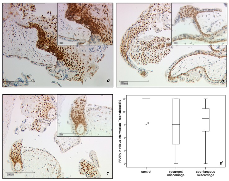
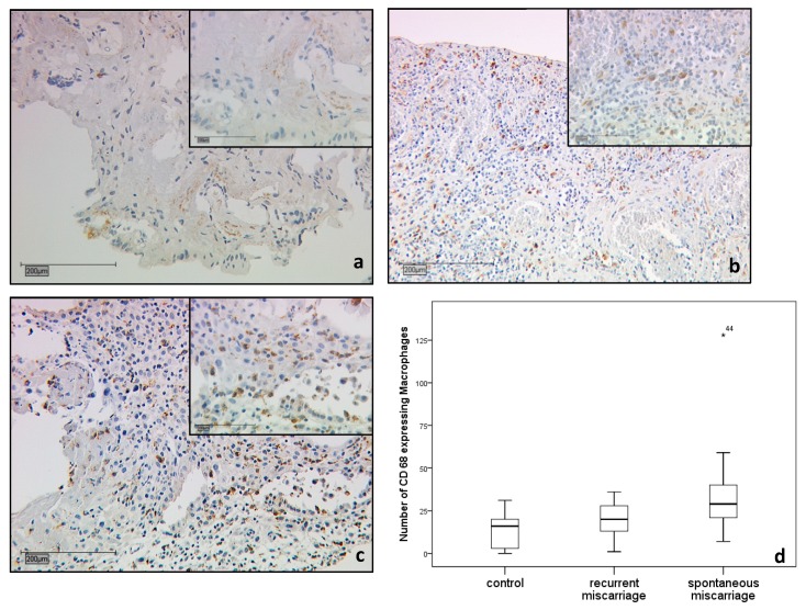
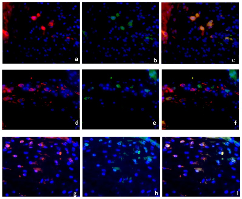
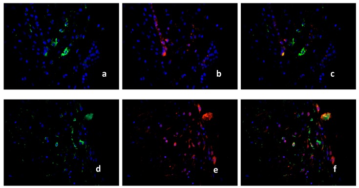
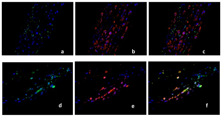
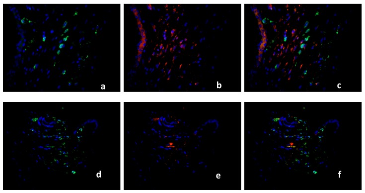
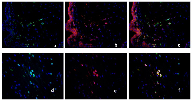
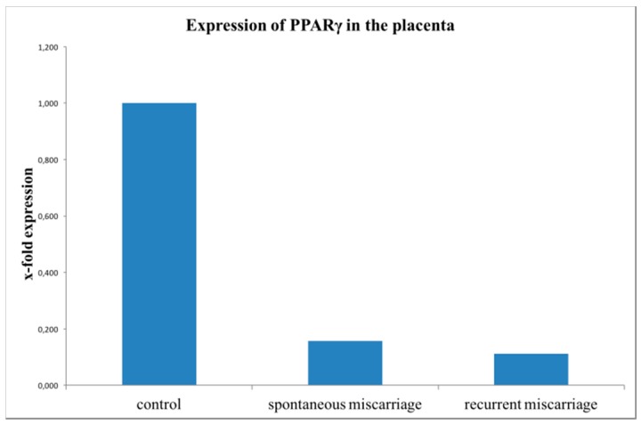
PPARγ belongs to the group of nuclear receptors which is expressed in the trophoblast and together with other factors is responsible for the maintenance of pregnancy. Apart from that PPARγ is also a main factor for macrophage polarization. The aim of this study was to investigate the combined expression pattern and frequency of PPARγ under physiological circumstances and in spontaneous and recurrent miscarriages in the trophoblast and in maternal macrophages of the decidua. Human placental tissues of the first trimester (15 physiologic pregnancies, 15 spontaneous abortion and 16 recurrent miscarriage placentas) were analyzed for expression of the nuclear receptor PPARγ. Expression changes were evaluated by immunohistochemistry and real time PCR (RT-PCR) in trophoblast and in maternal macrophages of the decidua. Maternal macrophages were identified by double immunofluorescence using cluster of differentiation 68 (CD68) as marker for macrophages and further characterized regarding their M1/M2 polarization status. The intermediate villous trophoblast revealed a significantly lower PPARγ expression in spontaneous and recurrent abortion. Maternal macrophages express PPARγ. Their number is significantly enhanced in the decidua of spontaneous miscarriages whereas in recurrent miscarriages maternal macrophages seem to express PPARγ only in very few cases. PPARγ is associated with an M2 polarization state that is common for decidual macrophages. The lack of PPARγ in recurrent miscarriage decidual macrophages seems to be associated with a specific inflammatory response against the fetus.

Keywords: PPARγ; decidual macrophages; first trimester placenta; miscarriage.


Conflict of interest statement

The authors declare no conflict of interest.

Figures

Figure 1
Figure 1
Immunohistochemical staining of Peroxisome proliferator-activated receptor gamma (PPARγ) in the villous trophoblast. PPARγ expression is found with high distribution and intensity in intermediate villous trophoblastic cells (IVT) of first-trimester placentas. The control group (15 cases) showed the strongest expression pattern (a). PPARγ was significantly downregulated (p = 0.01) in trophoblastic tissue of recurrent miscarriage (RM, 16 cases, (b). PPARγ expression in the IVT of spontaneous miscarriage tissue (SM, 15 cases), (c) was significantly decreased compared to the control (p = 0.001). The boxplot summarizes the statistical data of the immunohistochemical staining results (d). Scale is 200 μm. The insert picture is 100 μm scaled.
Figure 2
Figure 2
Immunohistochemical staining of decidual macrophages with CD68 as a marker for macrophage positivity. Decidual macrophages were increased in RM (16 cases) and SM samples (15 cases) compared to the control group (15 cases), (a). In recurrent miscarriage specimens, the decidual macrophages tended to be upregulated (p = 0.181) (b). In spontaneous miscarriage samples, the population of macrophages was significantly higher compared to the control (p = 0.013) (c). Summary of staining results of CD68 positive decidual macrophages (d). Scale is 200 μm. The insert picture is 100 μm scaled.
Figure 3
Figure 3
Double Immunofluorescence of CD68 & PPARγ. CD68 (red staining), PPARγ (green staining), nuclear staining (blue). CD68 (a) and PPARγ (b) are expressed in the decidua of healthy placenta (15 cases), co-expression presented as triple filter yellow (c). High distribution of CD68 positive macrophages was found in RM (16 cases), (d). PPARγ-positive cells are shown in (e). Triple filter excitation showed an absence of PPARγ positive macrophages (f). Both markers (CD68, (g) and PPARγ, (h)) are co-expressed in the group of SM (15 cases), (i). All pictures are 40× lens.
Figure 4
Figure 4
Double Immunofluorescence of CD68 and iNOS. CD68 (green staining), iNOS (red staining), nuclear staining (blue). CD68 (a) and iNOS (b) are co-expressed only in few macrophages in the decidua of healthy placenta, presented as triple filter yellow (c). High distribution of CD68 positive macrophages was found in RM (d). iNOS positive cells are shown in (e). Triple filter excitation shows a co-expression of iNOS and macrophages (f). All pictures are 40× lens.
Figure 5
Figure 5
Double Immunofluorescence of CD68 and TLR2. CD68 (green staining), TLR2 showed strong expression in the healthy decidua (red staining), nuclear staining (blue). CD68 (a) and TLR2 (b) are not co-expressed in the decidua of healthy placenta, presented as triple filter yellow (c). CD68 positive macrophages in RM placenta (d). TLR2 positive cells are shown in (e). Triple filter excitation shows a co-expression of TLR2 and macrophages (f) in the RM placenta. All pictures are 40× lens.
Figure 6
Figure 6
Double Immunofluorescence of CD68 and CCL1. CD68 (green staining), CCL1 showed intense expression in the healthy decidua (red staining), nuclear staining (blue). CD68 (a) and CCL1 (b) are co-expressed in the decidua of healthy placenta in a number of cells, presented as triple filter yellow (c). CD68 positive macrophages in RM placenta (d). Only few CCL1 positive cells are shown in (e). Triple filter excitation shows a diminished co-expression of CCL1 and macrophages (f) in the RM placenta. All pictures are 40× lens.
Figure 7
Figure 7
Double Immunofluorescence of CD68 and CX3CR1. CD68 (green staining), CX3CR1 showed expression in a variety of different cell types including endometrial glands and decidual stromal cells [21] in the healthy decidua (red staining), nuclear staining (blue). CD68 (a) and CX3CR1 (b) are co-expressed in the decidua of healthy placenta in almost all CD 68-positive cells (>95%), presented as triple filter yellow (c). CD68 positive macrophages in RM placenta (d). Only 40–50% CX3CR1 expressing cells (e) showed co-expression with CD68, as shown in (f). All pictures are 40× lens.
Figure 8
Figure 8
Results of PPARγ mRNA expression analysis with TaqMan RT-PCR from trophoblastic tissue. PPARγ mRNA expression was significantly downregulated in the miscarriage groups (SM, 15 cases, p = 0.01) and RM, 16 cases, p = 0.004)) compared to the healthy controls (15 cases). This bar graph shows the mean of relative PPARγ expression; therefore, the presentation of error bars is not appropriate.

Similar articles

Cited by

References

    1. Makrigiannakis A., Petsas G., Toth B., Relakis K., Jeschke U. Recent advances in understanding immunology of reproductive failure. J. Reprod. Immunol. 2011;90:96–104. doi: 10.1016/j.jri.2011.03.006. - DOI - PubMed
    1. Knabl J., Vattai A., Huttenbrenner R., Hutter S., Karsten M., Jeschke U. Rxralpha is upregulated in first trimester endometrial glands of spontaneous abortions unlike LXR and PPARγ. Eur. J. Histochem. 2016;60:2665. doi: 10.4081/ejh.2016.2665. - DOI - PMC - PubMed
    1. Toth B., Jeschke U., Rogenhofer N., Scholz C., Wurfel W., Thaler C.J., Makrigiannakis A. Recurrent miscarriage: Current concepts in diagnosis and treatment. J. Reprod. Immunol. 2010;85:25–32. doi: 10.1016/j.jri.2009.12.006. - DOI - PubMed
    1. Arck P.C., Rucke M., Rose M., Szekeres-Bartho J., Douglas A.J., Pritsch M., Blois S.M., Pincus M.K., Barenstrauch N., Dudenhausen J.W., et al. Early risk factors for miscarriage: A prospective cohort study in pregnant women. Reprod. Biomed. Online. 2008;17:101–113. doi: 10.1016/S1472-6483(10)60300-8. - DOI - PubMed
    1. Pestka A., Fitzgerald J.S., Toth B., Markert U.R., Jeschke U. Nuclear hormone receptors and female reproduction. Curr. Mol. Med. 2013;13:1066–1078. doi: 10.2174/1566524011313070002. - DOI - PubMed

MeSH terms

LinkOut - more resources