. 2018 Jun 26;7(7):66.
doi: 10.3390/cells7070066.

The Desmosomal Cadherin Desmoglein-2 Experiences Mechanical Tension as Demonstrated by a FRET-Based Tension Biosensor Expressed in Living Cells

Affiliations

The Desmosomal Cadherin Desmoglein-2 Experiences Mechanical Tension as Demonstrated by a FRET-Based Tension Biosensor Expressed in Living Cells

Sindora R Baddam et al. Cells. .

Abstract

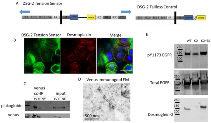
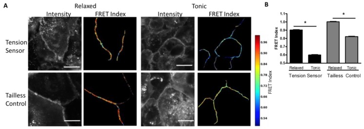
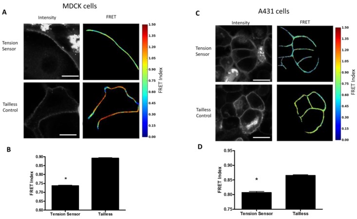
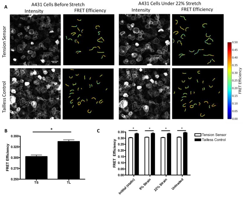
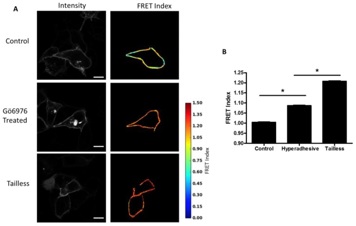
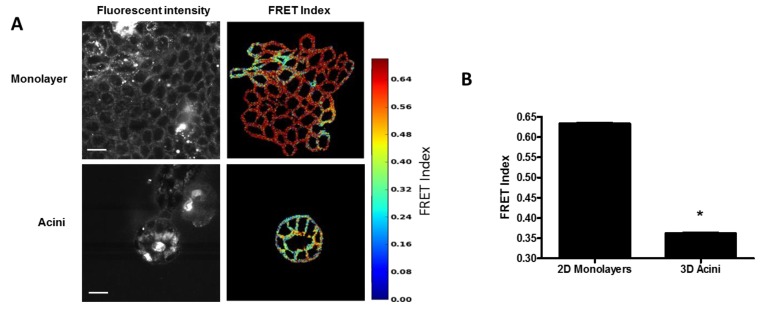
Cell-cell junctions are critical structures in a number of tissues for mechanically coupling cells together, cell-to-cell signaling, and establishing a barrier. In many tissues, desmosomes are an important component of cell-cell junctions. Loss or impairment of desmosomes presents with clinical phenotypes in the heart and skin as cardiac arrhythmias and skin blistering, respectively. Because heart and skin are tissues that are subject to large mechanical stresses, we hypothesized that desmosomes, similar to adherens junctions, would also experience significant tensile loading. To directly measure mechanical forces across desmosomes, we developed and validated a desmoglein-2 (DSG-2) force sensor, using the existing TSmod Förster resonance energy transfer (FRET) force biosensor. When expressed in human cardiomyocytes, the force sensor reported high tensile loading of DSG-2 during contraction. Additionally, when expressed in Madin-Darby canine kidney (MDCK) epithelial or epidermal (A431) monolayers, the sensor also reported tensile loading. Finally, we observed higher DSG-2 forces in 3D MDCK acini when compared to 2D monolayers. Taken together, our results show that desmosomes experience low levels of mechanical tension in resting cells, with significantly higher forces during active loading.

Keywords: cell biophysics; desmosomes; mechanobiology.


Conflict of interest statement

The authors declare no conflict of interest.

Figures

Figure 1
Figure 1
Design and validation of the desmoglein-2 (DSG-2) Tension Sensor. (A) Schematic for how TSmod was inserted into DSG-2 between the intracellular anchor (IA) and intracellular cadherin-type sequence (ICS) domains to make the DSG-2 tension sensor (other domains are EC, extracellular domain; EA, extracellular anchor; IPL, intracellular proline-rich linker; RUD, repeated unit domain; DTD, desmoglein-specific terminal domain). Under mechanical loading, the Förster resonance energy transfer (FRET) pair in TSmod is separated, reducing FRET. A tailless control was made by removing the cytoplasmic tail downstream of the tension sensor. The TSmod in the tailless control cannot be subjected to tensile load and, consequently, reports maximal FRET for the sensor. (B) The DSG-2 tension sensor expressed in A431 localized to cell-cell junctions. Arrows indicate co-localization with desmoplakin. Scale bar is 20 microns. (C) The DSG-2 tension sensor and DSG-2 tailless control were expressed in Madin-Darby canine kidney (MDCK) cells. Co-immunoprecipitation was performed with an antibody recognizing venus. Plakoglobin immunoprecipitated with the DSG-2 tension sensor, but not DSG-2 tailless. (D) MDCK cells expressing the DSG-2 tension sensor were fixed and processed for immunogold electron microscopy with anti-GFP antibody. A positive signal was observed at desmosomes, indicating that the DSG-2 tension sensor was localized to desmosomes. Scale bar is 500 nm. (E) Wild-type, DSG-2 CRISPR-cas9 knockout, and DSG-2 knockout reconstituted with DSG-2 tension sensor A431 cells were lysed and assessed for phosphorylated epidermal growth factor receptor (EGFR) (pY1173). Reconstitution with the DSG-2 tension sensor rescued the loss of phosphorylated EGFR seen in the knockout cells.
Figure 2
Figure 2
DSG-2 tensile force is increased in contracting cardiomyocytes. (A) Human iPSC-derived cardiomyocytes expressing the DSG-2 tension sensor or DSG-2 tailless control were exposed to either relaxing buffer (with actomyosin inhibitor 2,3-butanedione monoxime (BDM)) or tonic buffer (high K+). Tonic buffer resulted in a large decrease in FRET for the DSG-2 tension sensor. Tonic buffer also resulted in a small, but significant, decrease in FRET for the DSG-2 tailless control. Scale bar is 20 microns. (B) Quantification of FRET differences (representative experiment of three experiments with similar results, ANOVA with Newman-Keuls post-hoc test, * p < 0.001).
Figure 3
Figure 3
DSG-2 is under tension in resting epithelial cells. (A) MDCK cells expressing the DSG-2 tension sensor had reduced FRET compared to MDCK cells expressing the DSG-2 tailless control. Scale bar is 20 microns. (B) Quantification of FRET differences (representative experiment of five experiments with similar results, ANOVA with Newman-Keuls post-hoc test * p < 0.001). (C) A431 cells expressing the DSG-2 tension sensor had reduced FRET compared to A431 cells expressing the DSG-2 tailless control. Scale bar is 20 microns. (D) Quantification of FRET differences (representative experiment of three experiments with similar results, ANOVA with Newman-Keuls post-hoc test * p < 0.001).
Figure 4
Figure 4
FRET efficiency measurements of A431 cells. (A) FRET efficiency measurements of selected junctions from a monolayer of A431 cells expressing the DSG-2 tension sensor or tailless control. Scale bar is 50 microns. (B) Quantification of FRET efficiency measurements between tension sensor and tailless cells, differences were significant (ANOVA with Newman-Keuls post-hoc test, * p < 0.001). (C) DSG-2 Tension sensor and tailless expressing cells were subjected to 0% (static), 9%, and 22% strain, followed by an unloading. Images were acquired for the same cells at each condition (paired). No changes in FRET were observed by the stretch.
Figure 5
Figure 5
DSG-2 force is reduced in hyperadhesive cells. (A) MDCK cells expressing DSG-2 were treated with 10 nM of the protein kinase C (PKC) inhibitor, Gö6976, for 60 min to induce hyperadhesion. Cells stimulated with Gö6976 had increased FRET compared to control cells, indicating decreased tension. Scale bar is 20 microns. (B) Quantification of FRET differences (representative experiment of three experiments with similar results, ANOVA with Newman-Keuls post-hoc test, * p < 0.01).
Figure 6
Figure 6
Desmosome forces are increased in epithelial acini. (A) MDCK cells expressing the DSG-2 tension sensor were either seeded in Matrigel for seven to ten days to form acini or on top of thin Matrigel to form monolayers. Scale bar is 20 microns. (B) Quantification of FRET differences showed acini have an increased DSG-2 force compared to monolayers (representative experiment of three experiments with similar results, ANOVA with Newman-Keuls post-hoc test, * p < 0.01).

References

    1. Liu Z., Tan J.L., Cohen D.M., Yang M.T., Sniadecki N.J., Ruiz S.A., Nelson C.M., Chen C.S. Mechanical tugging force regulates the size of cell-cell junctions. Proc. Natl. Acad. Sci. USA. 2010;107:9944–9949. doi: 10.1073/pnas.0914547107. - DOI - PMC - PubMed
    1. Maruthamuthu V., Sabass B., Schwarz U.S., Gardel M.L. Cell-ECM traction force modulates endogenous tension at cell-cell contacts. Proc. Natl. Acad. Sci. USA. 2011;108:4708–4713. doi: 10.1073/pnas.1011123108. - DOI - PMC - PubMed
    1. Conway D.E., Breckenridge M.T., Hinde E., Gratton E., Chen C.S., Schwartz M.A. Fluid Shear Stress on Endothelial Cells Modulates Mechanical Tension across VE-Cadherin and PECAM-1. Curr. Biol. 2013;23:1024–1030. doi: 10.1016/j.cub.2013.04.049. - DOI - PMC - PubMed
    1. Borghi N., Sorokina M., Shcherbakova O.G., Weis W.I., Pruitt B.L., Nelson W.J., Dunn A.R. E-cadherin is under constitutive actomyosin-generated tension that is increased at cell-cell contacts upon externally applied stretch. Proc. Natl. Acad. Sci. USA. 2012;109:12568–12573. doi: 10.1073/pnas.1204390109. - DOI - PMC - PubMed
    1. Kottke M.D., Delva E., Kowalczyk A.P. The desmosome: Cell science lessons from human diseases. J. Cell Sci. 2006;119:797–806. doi: 10.1242/jcs.02888. - DOI - PubMed

LinkOut - more resources