Review
. 2018 Jun 26;7(7):67.
doi: 10.3390/cells7070067.

Rapid Evolution of Sperm Produces Diverse Centriole Structures that Reveal the Most Rudimentary Structure Needed for Function

Affiliations
Review

Rapid Evolution of Sperm Produces Diverse Centriole Structures that Reveal the Most Rudimentary Structure Needed for Function

Tomer Avidor-Reiss. Cells. .

Abstract

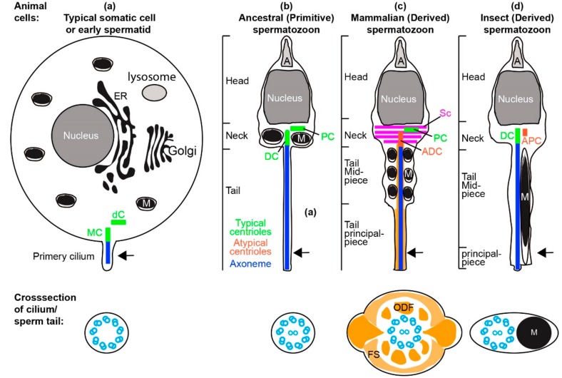
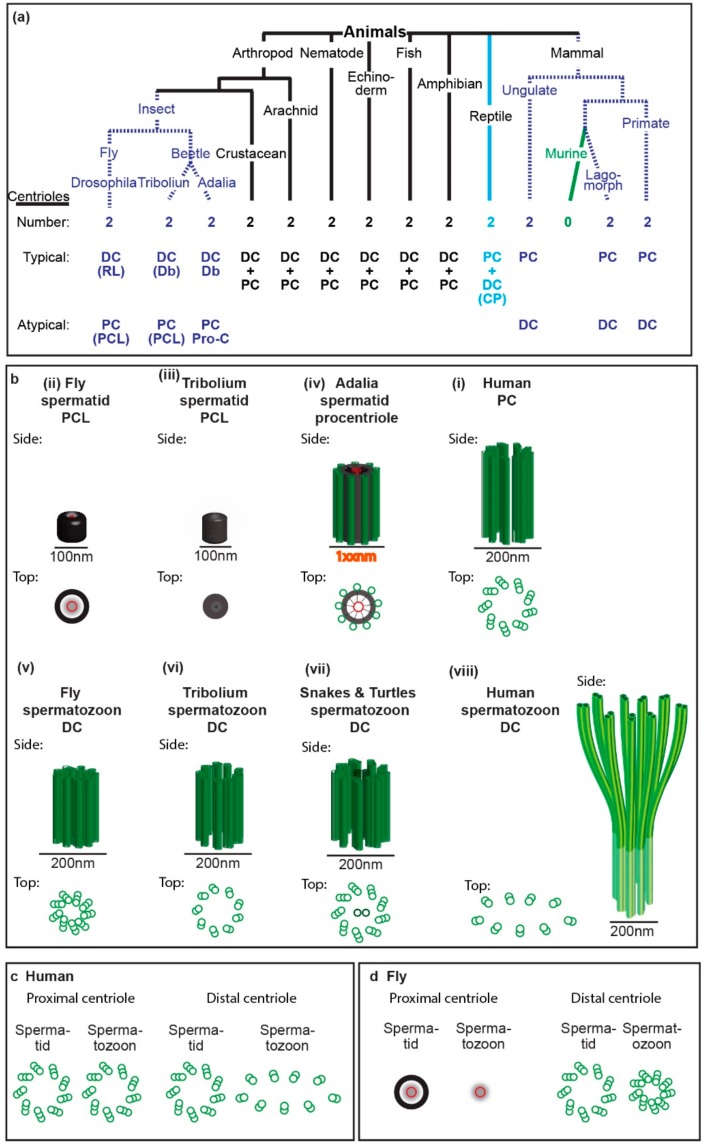
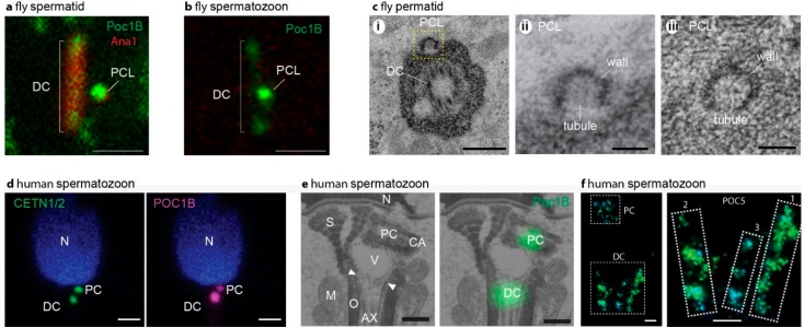
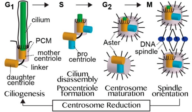
Centrioles are ancient subcellular protein-based organelles that maintain a conserved number and structure across many groups of eukaryotes. Centriole number (two per cells) is tightly regulated; each pre-existing centriole nucleates only one centriole as the cell prepares for division. The structure of centrioles is barrel-shaped, with a nine-fold symmetry of microtubules. This organization of microtubules is essential for the ancestral function of centriole⁻cilium nucleation. In animal cells, centrioles have gained an additional role: recruiting pericentriolar material (PCM) to form a centrosome. Therefore, it is striking that in animal spermatozoa, the centrioles have a remarkable diversity of structures, where some are so anomalous that they are referred to as atypical centrioles and are barely recognizable. The atypical centriole maintains the ability to form a centrosome and nucleate a new centriole, and therefore reveals the most rudimentary structure that is needed for centriole function. However, the atypical centriole appears to be incapable of forming a cilium. Here, we propose that the diversity in sperm centriole structure is due to rapid evolution in the shape of the spermatozoa head and neck. The enhanced diversity may be driven by a combination of direct selection for novel centriole functions and pleiotropy, which eliminates centriole properties that are dispensable in the spermatozoa function.

Keywords: centriole; centriole remodeling; centrosome; centrosome reduction; cilium; evolution; sperm; spermatogenesis.


Conflict of interest statement

The Authors declare no Competing Financial or Non-Financial Interests.

Figures

Figure 1
Figure 1
Centrioles in somatic cell and spermatozoa. (a) A representative undifferentiated somatic cell (e.g., stem cell in G1 phase of the cell cycle) or early spermatid with all organelles, and two typical centrioles: the mother centriole (MC) and daughter centriole (dC), and a short non-motile primary cilium. The primary cilium contains an axoneme made of nine doublets of microtubules. (b) Model of the ancestral “primitive” spermatozoa made of head, neck, and tail. The neck contains mitochondria (M) and two typical centrioles: the distal centriole (DC) and proximal centriole (PC). The Golgi forms the acrosome (A), the nucleus reshapes, and the endoplasmic reticulum (ER) is dramatically modified to a remnant. The sperm tail contains an axoneme made of nine doublets of microtubules with two central singlet microtubules. (c) Model of the mammalian spermatozoa made of head, neck, and tail that are divided into a midpiece and principle piece. The neck contains one typical proximal centriole and one atypical distal centriole. Additionally, the neck contains accessory structures that exhibit bilateral symmetry (e.g., striated column, SC). The mitochondria are found with the cytoplasmic axoneme in the midpiece [13]. The sperm tail contains the axoneme made of nine doublets of microtubules with two central singlet microtubules and accessory structures (outer dense fiber, ODF, and fibrous sheath, FS) that exhibit bilateral symmetry. (d) Model of the insect spermatozoa made of head, neck, and tail that is mostly a midpiece and with a tiny principle piece. The neck contains one typical distal centriole and one atypical proximal centriole. The sperm tail contains the axoneme made of nine doublets of microtubules with two central singlet microtubules. Asymmetrically located mitochondria are found along one side of the cytoplasmic axoneme along most of the sperm tail; this section is homologues to the midpiece in mammalian sperm [14]. Arrow in the animal cells indicated the plane of the cross-section in the cilium/sperm tail.
Figure 2
Figure 2
Spermatozoon typical and atypical centrioles have diverse structures in animals. (a) Phylogenetic tree with representative animal groups. Number of centrioles (first row), which centriole is typical and atypical (second and third rows, respectively), and type of centrioles as well as their modifications (in parenthesis) are indicated; RL, reduced diameter of the centriole lumen; Db, nine doublets of microtubules instead of triplets; PCL, proximal centriole-like structure as depicted in panel “b” or “d”; Pro-c, procentriole; CP, centriole with central pair. (b) Models of spermatid and spermatozoa proximal and distal centriole. The centrioles are depicted from side and top views and are to scale. (c,d) The structural differences between spermatid centrioles and spermatozoa centrioles in human (c) and fly (d).
Figure 3
Figure 3
Sperm centrioles in flies (ac) and mammals (df). (a) Fluorescent microscope picture of fly spermatid with DC and PCL labeled by the centriolar protein Poc1B and Ana1. (b) Fluorescent microscope picture of fly spermatozoon with Poc1B faintly labeling the DC and intensely labeling the PCL. In (a,b) the scale bar is 1 μm, Poc1B is genetically tagged by GFP, and Ana1 is genetically tagged by tdTomato. (c) Electron microscope picture of fly spermatozoon centrioles. (i) A section in a plane that has a cross-section of the DC and longitudinal section of the PCL (yellow box). (ii) Zoom in the PCL boxed in (i). (iii) A cross-section of the PCL depicting the wall and central tubule. In (c), the scale bar is 0.1 μm. (d) Fluorescent microscope picture of a human spermatozoon with CENTE1/2 or POC1B labeling the DC and PCL. The scale bar is 1 μm. CENTE1/2 and Poc1B are labeled by specific antibodies. (e) Correlative light and electron microscopy picture of human spermatozoon centrioles. On the left, electron microscopy section depicting POC1B labeling of the PC that is found near the nucleus, and the DC that is attached to the axoneme. Ax, axoneme; M, mitochondria; mp, tail midpiece; N, nucleus; ne, neck; O, outer dance fibers; S, striated columns; V; vault; CA; centriole adjunct. (f) Stochastic Optical Reconstruction Microscopy (STORM) super-resolution fluorescent microscopy picture of bovine spermatozoon centrioles. On the left, a picture depicting POC5 labeling of the PC and the DC. On the right, zooming in on the DC identifies two major rods (marked as “1” and “2”) and one minor rod (marked as “3”) labeled by POC5. In (f), the scale bar is 0.1 μm. Panels (ac) are from Khire 2016 [15]. Panels (df) are from Fishman [9].
Figure 4
Figure 4
A model of the centriole cycle. Cells have two centrioles: the mother centriole and daughter centriole. The two centrioles have distinct roles in controlling cilium number, and in some cell types, in the outcome of asymmetric cell division. The mother centriole is the older of the two centrioles and is compositionally and structurally fully developed (mature). However, its function differs depending upon the present stage of the cell cycle. When a cell is in G1 phase, the mother centriole is enveloped by a thin layer of pericentriolar material (PCM) and assembles a cilium. In S phase, the cilium disassembles and the mother centriole forms a new centriole (the procentriole). In G2 phase, the mother centriole recruits the PCM, which nucleates and anchors microtubules to form an aster. In M phase, the mother centriole participates in the formation and localization of one of the mitotic spindle poles. After the completion of cell division, each centriole pair becomes a mother and a daughter centriole for one of the two daughter cells, and the mother centriole loses most of its PCM and astral microtubules through a process known as centrosome reduction. The daughter centriole is the younger of the two centrioles, and is compositionally, structurally, and functionally immature. When the cell is in S phase, the daughter centriole forms a procentriole. In G2 phase, the daughter separates from its mother, recruits PCM, forms an aster, and then forms the second centrosome. In M phase, the daughter centriole participates in the formation and localization of the second mitotic spindle pole, and after cell division becomes a mother centriole for one of the daughter cells.
Figure 5
Figure 5
Mechanisms of atypical centriole formation in mammals and insects. (a) Model of centriole remodeling that transforms the typical centriole to an atypical centriole, and the role of sperm centrioles in the zygotes of non-rodent mammals. The early spermatid centrosome is made of two typical centrioles (PC and DC) and pericentriolar material (PCM) that form an aster. During spermiogenesis, centrosomal proteins are redistributed, and their amount is altered (reduced or enriched). Consequently, the DC is remodeled and forms an atypical centriole: the spermatozoon distal centriole (SDC). The SDC is made of rods and splayed microtubules (mts), which are attached to the core of the flagellum (axoneme, Ax) in the mature spermatozoon. The PC remains structurally typical, but it has an atypical protein composition. After fertilization, both the typical and atypical centrioles recruit maternal PCM and reconstitute the zygote centrosome. Later, these typical and atypical centrioles separate and each form a centrosome, an aster, a new centriole (procentriole), and a spindle pole at distinct stages of zygote development. (b) Model of atypical centriole formation, followed by centriole remodeling, and the role of sperm centrioles in the zygote of insect. The early spermatid centrosome consists of a single centriole that becomes distal centriole (DC). The DC is surrounded by PCM and forms an aster. During early spermiogenesis, an atypical centriole forms: the PCL. During mid and late spermiogenesis, centrosomal proteins are redistributed, and their amount is altered (reduced or enriched). Consequently, the DC protein composition is remodeled, but it maintains a centriole wall made of microtubules, which are attached to the core of the flagellum (axoneme, Ax) in the mature spermatozoon. The PCL is modified structurally and compositionally. After fertilization, both the DC and PCL recruit maternal PCM and reconstitute the zygote centrosome, which later forms an aster, a new centriole (procentriole), and a spindle pole at distinct stages of zygote development.

References

    1. Swann K., Lai F.A. Egg Activation at Fertilization by a Soluble Sperm Protein. Physiol. Rev. 2016;96:127–149. doi: 10.1152/physrev.00012.2015. - DOI - PubMed
    1. Ostermeier G.C., Miller D., Huntriss J.D., Diamond M.P., Krawetz S.A. Reproductive biology: Delivering spermatozoan RNA to the oocyte. Nature. 2004;429:154. doi: 10.1038/429154a. - DOI - PubMed
    1. Gur Y., Breitbart H. Mammalian sperm translate nuclear-encoded proteins by mitochondrial-type ribosomes. Genes Dev. 2006;20:411–416. doi: 10.1101/gad.367606. - DOI - PMC - PubMed
    1. Eberhard W.G. Postcopulatory sexual selection: Darwin’s omission and its consequences. Proc. Natl. Acad. Sci. USA. 2009;106:10025–10032. doi: 10.1073/pnas.0901217106. - DOI - PMC - PubMed
    1. Parker G.A., Pizzari T. Sperm competition and ejaculate economics. Biol. Rev. Camb. Philos. Soc. 2010;85:897–934. doi: 10.1111/j.1469-185X.2010.00140.x. - DOI - PubMed

LinkOut - more resources