. 2018 Aug 29;92(18):e00036-18.
doi: 10.1128/JVI.00036-18. Print 2018 Sep 15.

Tomato Yellow Leaf Curl Virus V2 Interacts with Host Histone Deacetylase 6 To Suppress Methylation-Mediated Transcriptional Gene Silencing in Plants

Affiliations

Tomato Yellow Leaf Curl Virus V2 Interacts with Host Histone Deacetylase 6 To Suppress Methylation-Mediated Transcriptional Gene Silencing in Plants

Bi Wang et al. J Virol. .

Abstract

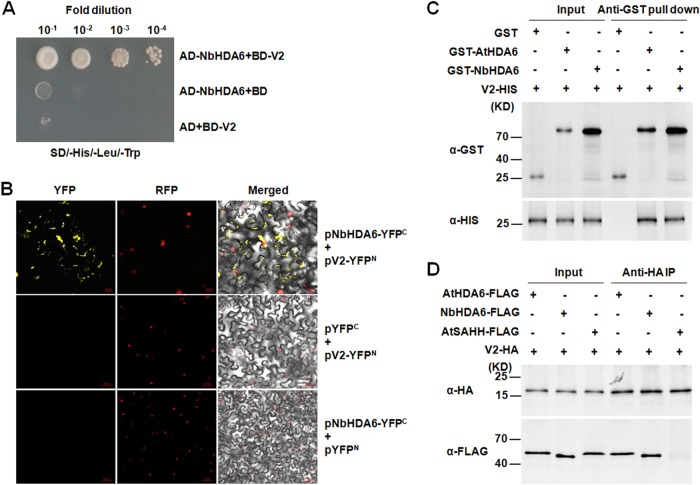
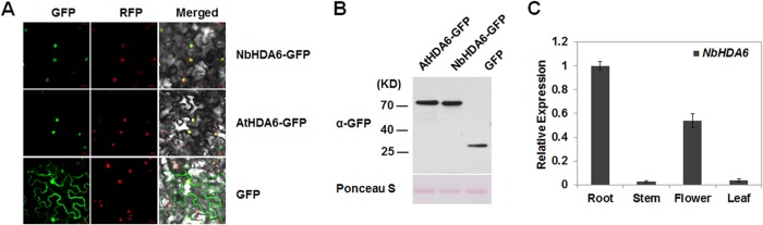
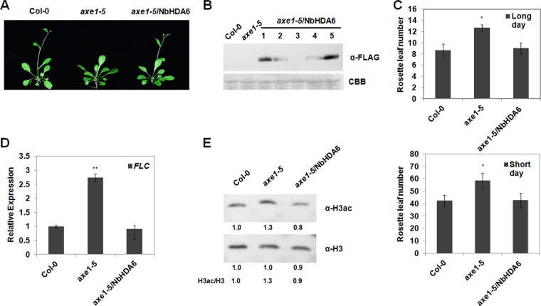
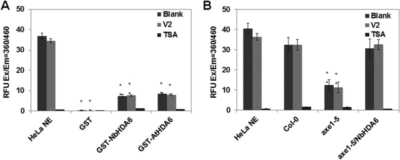
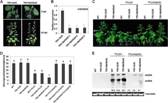
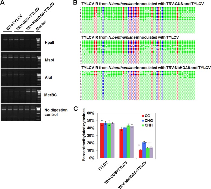
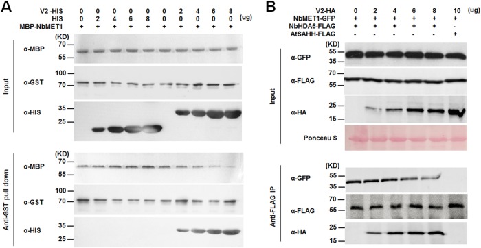
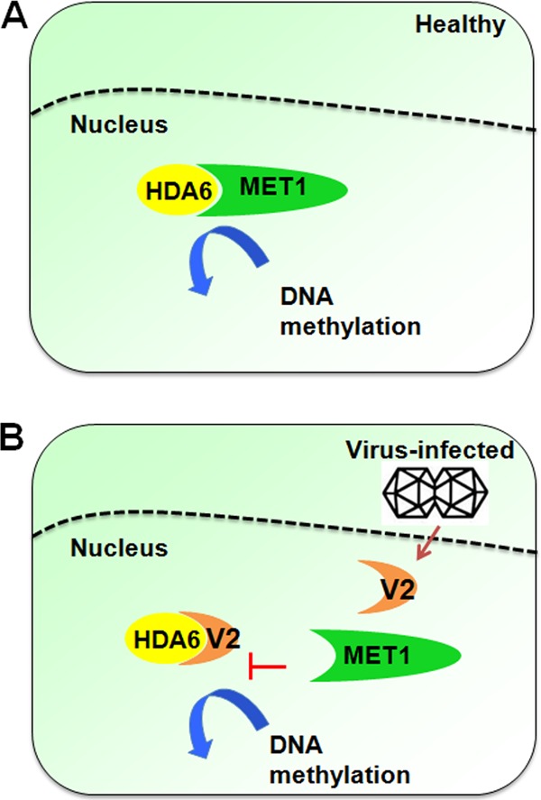
Cytosine DNA methylation is a conserved epigenetic silencing mechanism that defends against biotic stresses such as geminivirus infection. As a countermeasure, geminiviruses encode proteins that inhibit methylation and transcriptional gene silencing (TGS). Previous studies showed that V2 protein of Tomato yellow leaf curl virus (TYLCV) functions as a TGS suppressor. However, how V2 mediates TGS suppression remains unknown. Here we show that V2 interacts directly with a Nicotiana benthamiana histone deacetylase 6 (NbHDA6), a homolog of Arabidopsis HDA6 (AtHDA6), known to be involved in gene silencing in cooperation with methyltransferase 1 (MET1). NbHDA6 genetically complemented a late-flowering phenotype and restored histone deacetylation of an AtHDA6 mutant. Furthermore, our investigation showed that NbHDA6 displayed histone deacetylase enzymatic activity, which was not inhibited by V2. Genetic analysis revealed that silencing of NbHDA6 expression resulted in enhanced susceptibility to TYLCV infection. In addition, methylation-sensitive PCR and bisulfite sequencing analysis showed that silencing of NbHDA6 expression caused reduced DNA methylation of the viral genome in infected plants. HDA6 was previously shown to recruit and physically interact with MET1 to function in gene silencing. Using competitive pulldown and coimmunoprecipitation assays, we demonstrated that V2 did not interact but competed with NbMET1 for direct binding to NbHDA6. These findings suggest that V2 interacts with host HDA6 and interferes with the recruitment of MET1 by HDA6, resulting in decreased methylation of the viral DNA genome by TGS with a concomitant increase in host susceptibility to TYLCV infection.IMPORTANCE Plants employ repressive viral genome methylation as an epigenetic defense against geminiviruses. In turn, geminiviruses encode proteins that inhibit methylation by TGS. Previous studies showed that TYLCV V2 can efficiently suppress TGS, but the mechanism remains unknown. We showed that V2 interacted with NbHDA6 but did not inhibit its enzymatic activity. As HDA6 is known to be involved in gene silencing in cooperation with MET1, we explored the relationship between V2, NbMET1, and NbHDA6. Our investigation showed that V2 did not interact but competed with NbMET1 for direct binding to NbHDA6. To our knowledge, this is the first report that viral proteins inhibit TGS by interacting with histone deacetylase but not by blocking the methyl cycle. This work provides an additional mechanism for TGS suppression by geminiviruses.

Keywords: DNA methylation; MET1; Tomato yellow leaf curl virus; V2; geminivirus; histone deacetylase 6 (HDA6); methyltransferase 1; suppressor; suppressor of RNA silencing; transcriptional gene silencing (TGS).


Figures

FIG 1
FIG 1
TYLCV V2 physically interacts with NbHDA6. (A) Yeast two-hybrid assay between NbHDA6 and V2. Yeast strain Y2HGold cotransformed with the indicated plasmids was spotted on synthetic depleted (SD) growth medium lacking histidine, leucine, and tryptophan in 10-fold serial dilutions. (B) BiFC visualization of an interaction between V2 and NbHDA6 in N. benthamiana leaves. RFP-histone 2B (RFP-H2B) was used as a marker for the nucleus. Left panels, YFP fluorescence; middle panels, RFP fluorescence; right panels, YFP/RFP/bright-field overlay. Bars, 20 μm. (C) Pulldown assay for detecting an in vitro interaction between NbHDA6/AtHDA6 and V2. Equal molar amounts of GST or GST fusion proteins were used to pull down HIS fusion proteins. Immunoblot assays were performed using anti-HIS or anti-GST antibody to detect the associated proteins. (D) In vivo co-IP assay for the interaction between NbHDA6/AtHDA6 and V2. Approximately 1% of input and one-quarter of eluted protein complex were analyzed by immunoblotting. These experiments were repeated three times with similar results.
FIG 2
FIG 2
NbHDA6 localization and expression. (A) Subcellular localization of HDA6. Micrographs showing cells expressing NbHDA6-GFP, AtHDA6-GFP, or GFP were examined under GFP fluorescence (left panels), RFP fluorescence (middle panels), or an overlay of GFP/RFP/bright illumination (right panels) by confocal microscopy. RFP-H2B was used as a marker for the nucleus. Bars, 20 μm. (B) Western blot analysis to detect NbHDA6-GFP, AtHDA6-GFP, or GFP protein expression using anti-GFP antibody. The gel was stained with Ponceau S to show protein loadings. (C) Real-time RT-PCR analysis of NbHDA6 mRNA levels in N. benthamiana. GAPDH was used as an internal control. Each data set was derived from at least three biological repeats.
FIG 3
FIG 3
Genetic complementation of the Arabidopsis axe1-5 mutant by NbHDA6. (A) Comparison of growth phenotypes of the axe1-5 mutant, the complementation line axe1-5/NbHDA6, and Col-0 plants under long-day (LD) conditions. (B) Immunoblot assays were performed using anti-FLAG antibody to detect the NbHDA6 protein expression of axe1-5/NbHDA6 transgenic plants. Five independent axe1-5/NbHDA6 transgenic Arabidopsis lines (lanes 1 to 5) are shown. The gel was stained with Coomassie brilliant blue (CBB) to show protein loadings. Two lines (1 and 5) were selected for further characterization because of high expression levels. (C) Rosette leaf numbers at the initiation of flowering for axe1-5 and axe1-5/NbHDA6 transgenic plants grown under LD or short-day (SD) conditions compared with Col-0 plants. Student's t test was performed using rosette leaf numbers at the initiation of flowering from six individual plants. A single asterisk indicates a significant difference (P < 0.05) between the two-paired samples. (D) Real-time RT-PCR analysis of FLC expression in Col-0, axe1-5, and axe1-5/NbHDA6 plants. Actin was used as an internal control. Student's t test was performed, and double asterisks indicate a significant difference (P < 0.01) between the two-paired samples. (E) Western blot analysis to detect acetylated H3 (H3ac) using H3ac antibody in protein extracts from Col-0, axe1-5, and axe1-5/NbHDA6 transgenic lines. Total H3 protein was used as loading control. Each data set was derived from at least three biological repeats. Numbers below the blots indicate the relative abundances of H3ac or H3 proteins calculated by ImageJ.
FIG 4
FIG 4
Deacetylase activity of NbHDA6 is not affected by V2. (A) Detection of the histone deacetylase activity of GST-NbHDA6 and GST-AtHDA6 using an HDAC activity fluorometric assay kit. HDA6 mixed with large amounts of V2 (32-fold molar excess V2:HDA6) or HDAC inhibitor TSA were also assessed. HeLa nuclear extract (HeLa NE) served as the positive control, and purified GST protein served as the negative control. (B) Detection of the histone deacetylase activity of nuclear protein extracts from Col-0, axe1-5, and axe1-5/NbHDA6 plants using an HDAC activity fluorometric assay kit. Nuclear protein extracts mixed with large amounts of V2 or HDAC inhibitor TSA were also assessed. HeLa NE served as the positive control. The relative fluorescence units (RFU) generated from the assay for each mixture are compared. Error bars represent the standard errors of the means from independent measurements. Student's t test was performed, and double asterisks indicate a significant difference (P < 0.01) between the two-paired samples. Each data set was derived from at least three biological repeats.
FIG 5
FIG 5
N. benthamiana HDA6 silenced lines show enhanced susceptibility to TYLCV infection. (A) Comparison of phenotypes of N. benthamiana plants in which the NbHDA6 gene was silenced (TRV-NbHDA6) and those of plants treated with a control silencing vector (TRV-GUS). (B) Relative NbHDA6 gene expression levels in VIGS and control N. benthamiana determined by real-time RT-PCR. TRV-NbHDA6-1, TRV-NbHDA6-2, and TRV-NbHDA6-3 represented three independent VIGS plants, and TRV-GUS represents the control plants. GAPDH was used as an internal control. Student's t test was performed, and double asterisks indicate a significant difference (P < 0.01) between the two-paired samples. Each data set was derived from at least three biological repeats. (C) Phenotypes of TYLCV or V2-knockout TYLCV [TYLCV(ΔV2)]-infected wild-type, TRV-GUS control, and NbHDA6-silenced plants. Leaves were photographed 30 days after inoculation. (D) Aboveground plant heights of TYLCV- or TYLCV(ΔV2)-infected WT, TRV-GUS, and NbHDA6-silenced plants. Means with the same letter are not significantly different (SPSS, P = 0.05). (E) Southern blot analysis of TYLCV viral DNA accumulation in systemic leaves of N. benthamiana infected with TYLCV or TYLCV(ΔV2). The DNA agarose gel was stained with ethidium bromide for genomic DNA as loading control and then blotted using probes specific for TYLCV. Numbers below the blot indicated relative abundances of TYLCV viral DNA calculated by ImageJ. Similar results were observed in at least three independent experiments.
FIG 6
FIG 6
NbHDA6 positively regulated the cytosine methylation of TYLCV. (A) Analysis of DNA methylation of the TYLCV genome by methylation-sensitive PCR. Genomic DNA was digested with HpaII, MspI, AluI, or McrBC and then loaded into the PCR system. Undigested DNA is shown as a control. (B) Cytosine methylation profiles assessed by bisulfite sequencing. The circles represent cytosine residues and are color coded according to the sequence context (red for CG, blue for CHG, and green for CHH). Solid circles indicate methylated cytosines. Each line represents the sequence of an individual clone. (C) Percentages of methylated cytosines in the TYLCV intergenic regions (IR). Student's t test was performed using the methylation values from individual clones. Double asterisks indicate a significant difference (P < 0.01) between the two-paired samples. Error bars represent the standard errors of the means from independent measurements. Samples were prepared by pooling six leaves from six systemically infected plants at 30 dpi. Similar results were observed in at least three independent experiments.
FIG 7
FIG 7
V2 competes with NbMET1 for direct binding to NbHDA6. (A) In vitro competitive pulldown assays. The indicated amounts of V2-HIS or HIS protein were mixed with 2 μg of MBP-NbMET1 and pulled down by 2 μg of GST-NbHDA6. The bound protein was detected by immunoblotting with the indicated antibodies. (B) In vivo competitive co-IP assays. NbHDA6-FLAG and NbMET1-GFP were coexpressed in Arabidopsis protoplasts in the presence or absence of V2-HA. The immune complexes were pulled down by using anti-FLAG agarose beads. These experiments were repeated three times with similar results.
FIG 8
FIG 8
Proposed model for the NbHDA6-V2 interaction in regulation of methylation-mediated TGS during geminivirus infection. (A) HDA6 is a conserved regulator for inactivating and constitutively silencing genomic regions through DNA methylation. HDA6 recruits and physically interacts with MET1 to function cooperatively in gene silencing. (B) The V2 protein encoded by TYLCV is a TGS suppressor. This model proposes that V2 targets NbHDA6, thereby interfering with the recruitment of MET1 by HDA6. This results in a decrease in methylation of the viral DNA, leading to an increase in host susceptibility to TYLCV infection.

Similar articles

Cited by

References

    1. Zhang X, Yazaki J, Sundaresan A, Cokus S, Chan SW, Chen H, Henderson IR, Shinn P, Pellegrini M, Jacobsen SE, Ecker JR. 2006. Genome-wide high-resolution mapping and functional analysis of DNA methylation in Arabidopsis. Cell 126:1189–1201. doi:10.1016/j.cell.2006.08.003. - DOI - PubMed
    1. Law JA, Jacobsen SE. 2010. Establishing, maintaining and modifying DNA methylation patterns in plants and animals. Nat Rev Genet 11:204–220. doi:10.1038/nrg2719. - DOI - PMC - PubMed
    1. Bisaro DM. 2006. Silencing suppression by geminivirus proteins. Virology 344:158–168. doi:10.1016/j.virol.2005.09.041. - DOI - PubMed
    1. Pooggin MM. 2013. How can plant DNA viruses evade siRNA-directed DNA methylation and silencing? Int J Mol Sci 14:15233–15259. doi:10.3390/ijms140815233. - DOI - PMC - PubMed
    1. Jackel JN, Storer JM, Coursey T, Bisaro DM. 2016. Arabidopsis RNA polymerases IV and V are required to establish H3K9 methylation, but not cytosine methylation, on geminivirus chromatin. J Virol 90:7529–7540. doi:10.1128/JVI.00656-16. - DOI - PMC - PubMed

Publication types

Supplementary concepts

LinkOut - more resources